Copyright
©The Author(s) 2015.
World J Clin Cases. Mar 16, 2015; 3(3): 206-209
Published online Mar 16, 2015. doi: 10.12998/wjcc.v3.i3.206
Published online Mar 16, 2015. doi: 10.12998/wjcc.v3.i3.206
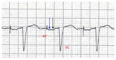
Figure 1 ECG after implant showing atrial-based pacing with spontaneous ventricular activation.
AP-VS interval: 280-300 msec. Right atrial stimulus artifact is followed by a first deflection corresponding to right atrial depolarization (blue arrow) and then a second corresponding to left atrial depolarization (black arrow). AP: Atrial pacing; VS: Ventricular sensing.
- Citation: Maria ED, Olaru A, Cappelli S. Minimizing right ventricular pacing in sinus node disease: Sometimes the cure is worse than the disease. World J Clin Cases 2015; 3(3): 206-209
- URL: https://www.wjgnet.com/2307-8960/full/v3/i3/206.htm
- DOI: https://dx.doi.org/10.12998/wjcc.v3.i3.206


