
















©The Author(s) 2015.
World J Clin Cases. Feb 16, 2015; 3(2): 191-195
Published online Feb 16, 2015. doi: 10.12998/wjcc.v3.i2.191
Published online Feb 16, 2015. doi: 10.12998/wjcc.v3.i2.191
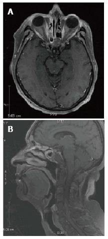
Figure 5 T1 magnetic resonance imaging 30 mo post-treatment.
A: Axial T1 magnetic resonance imaging (MRI) and B: Sagittal T1 MRI show no evidence of recurrent disease. There is a decrease of the previously noted inflammatory changes in the sinuses. There is retained fluid in the bilateral frontal and left sphenoid sinuses, without bony destruction or expansion.
- Citation: Noticewala SS, Mell LK, Olson SE, Read W. Survival in unresectable sinonasal undifferentiated carcinoma treated with concurrent intra-arterial cisplatin and radiation. World J Clin Cases 2015; 3(2): 191-195
- URL: https://www.wjgnet.com/2307-8960/full/v3/i2/191.htm
- DOI: https://dx.doi.org/10.12998/wjcc.v3.i2.191