Copyright
©The Author(s) 2015.
World J Clin Cases. Feb 16, 2015; 3(2): 191-195
Published online Feb 16, 2015. doi: 10.12998/wjcc.v3.i2.191
Published online Feb 16, 2015. doi: 10.12998/wjcc.v3.i2.191
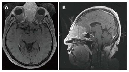
Figure 1 T1 magnetic resonance imaging of sinonasal undifferentiated carcinoma neoplasm prior to treatment.
A: Axial T1 magnetic resonance imaging (MRI) and B: Sagittal T1 MRI show an avidly enhancing mass centered in the left ethmoid air cells with extension into the left frontal sinus with adjacent retained fluid and maxillary sinus with erosion of the medial orbital walls bilaterally, left greater than right. The majority of the ethmoid air cells are replaced by the neoplasm. Extension through the cribriform plate is noted with involvement of the left olfactory lobe, predominantly along the gyrus rectus. There is extensive surrounding edema in the left frontal lobe, extending back to the frontal horn of the left lateral ventricle.
- Citation: Noticewala SS, Mell LK, Olson SE, Read W. Survival in unresectable sinonasal undifferentiated carcinoma treated with concurrent intra-arterial cisplatin and radiation. World J Clin Cases 2015; 3(2): 191-195
- URL: https://www.wjgnet.com/2307-8960/full/v3/i2/191.htm
- DOI: https://dx.doi.org/10.12998/wjcc.v3.i2.191


