Copyright
©The Author(s) 2015.
World J Clin Cases. Dec 16, 2015; 3(12): 1000-1004
Published online Dec 16, 2015. doi: 10.12998/wjcc.v3.i12.1000
Published online Dec 16, 2015. doi: 10.12998/wjcc.v3.i12.1000
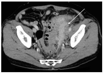
Figure 1 Follow-up computed tomography.
Computed tomography shows an ill-demarcated intrapelvic mass lesion extending to the left lower ureter, left margin of the bladder, and sigmoid colon, as denoted by arrow.
- Citation: Yabuuchi Y, Matsubayashi H, Matsuzaki M, Shiomi A, Moriguchi M, Kawamura I, Ito I, Ono H. Colovesical fistula caused by glucocorticoid therapy for IgG4-related intrapelvic mass. World J Clin Cases 2015; 3(12): 1000-1004
- URL: https://www.wjgnet.com/2307-8960/full/v3/i12/1000.htm
- DOI: https://dx.doi.org/10.12998/wjcc.v3.i12.1000


