
















©The Author(s) 2015.
World J Clin Cases. Jan 16, 2015; 3(1): 85-88
Published online Jan 16, 2015. doi: 10.12998/wjcc.v3.i1.85
Published online Jan 16, 2015. doi: 10.12998/wjcc.v3.i1.85
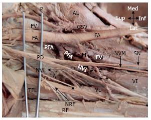
Figure 2 Right femoral region showing.
Med: Medial; Lat: Lateral; Sup: Superior; Inf: Inferior; FA: Femoral Artery; FV: Femoral Vein; GSV: Great Saphenous Vein; AL: Adductor longus; P: Pectineus; PFA: Profunda femoris artery; LCFA: Lateral circumflex femoral artery; PD: Posterior division of femoral nerve; M: Muscular branch of FA; TFL: Tensor fascia lata; RF: Rectus femoris; VI: Vastus intermedialis; VL: Vastus lateralis; NVM: Nerve to vastus medialis; SN: Saphenous nerve; NVL: Nerve to vastus lateralis; NVI: Nerve to vastus intermedius; NRF: Nerve to rectus femoris; #: Terminal branches of M; A: Ascending branch of LCFA; T: transverse branch of LCFA.
- Citation: Goel S, Arora J, Mehta V, Sharma M, Suri RK, Rath G. Unusual disposition of lateral circumflex femoral artery: Anatomical description and clinical implications. World J Clin Cases 2015; 3(1): 85-88
- URL: https://www.wjgnet.com/2307-8960/full/v3/i1/85.htm
- DOI: https://dx.doi.org/10.12998/wjcc.v3.i1.85