Copyright
©2014 Baishideng Publishing Group Inc.
World J Clin Cases. Aug 16, 2014; 2(8): 385-390
Published online Aug 16, 2014. doi: 10.12998/wjcc.v2.i8.385
Published online Aug 16, 2014. doi: 10.12998/wjcc.v2.i8.385
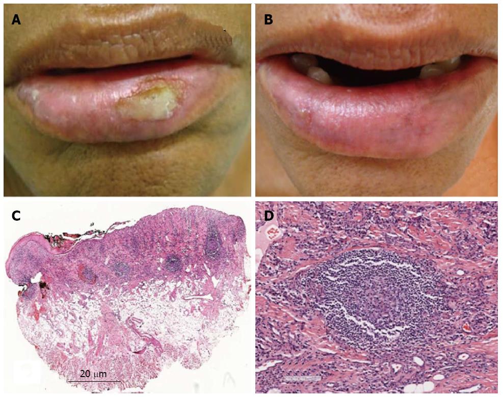
Figure 2 Case 2.
A: Clinical aspect at the first appointment, showing lower lip edema, dryness and ulcer on the left side of the semimucosa; B: Clinical aspect at the second appointment, showing remission of the ulceration on the left side, and only a small ulcer on the right side of the semimucosa; C: Histological aspects. Lower power view showing epithelial atrophy and ulceration. In the connective tissue, an intense, diffuse inflammatory infiltrate extending deep into the fatty tissue, with some lymphoid follicles was observed (HE); D: Secondary lymphoid follicle (HE).
- Citation: Miranda AM, Ferrari TM, Werneck JT, Junior AS, Cunha KS, Dias EP. Actinic prurigo of the lip: Two case reports. World J Clin Cases 2014; 2(8): 385-390
- URL: https://www.wjgnet.com/2307-8960/full/v2/i8/385.htm
- DOI: https://dx.doi.org/10.12998/wjcc.v2.i8.385


