
















©2014 Baishideng Publishing Group Inc.
World J Clin Cases. Jul 16, 2014; 2(7): 284-288
Published online Jul 16, 2014. doi: 10.12998/wjcc.v2.i7.284
Published online Jul 16, 2014. doi: 10.12998/wjcc.v2.i7.284
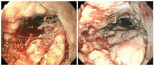
Figure 1 Upper endoscopy showing an irregular velvety appearing intraluminal mass in the esophagus (A and B).
- Citation: Ramani C, Shah N, Nathan RS. Verrucous carcinoma of the esophagus: A case report and literature review. World J Clin Cases 2014; 2(7): 284-288
- URL: https://www.wjgnet.com/2307-8960/full/v2/i7/284.htm
- DOI: https://dx.doi.org/10.12998/wjcc.v2.i7.284