
















Copyright: ©Author(s) 2026.
World J Clin Cases. Mar 26, 2026; 14(9): 118634
Published online Mar 26, 2026. doi: 10.12998/wjcc.v14.i9.118634
Published online Mar 26, 2026. doi: 10.12998/wjcc.v14.i9.118634
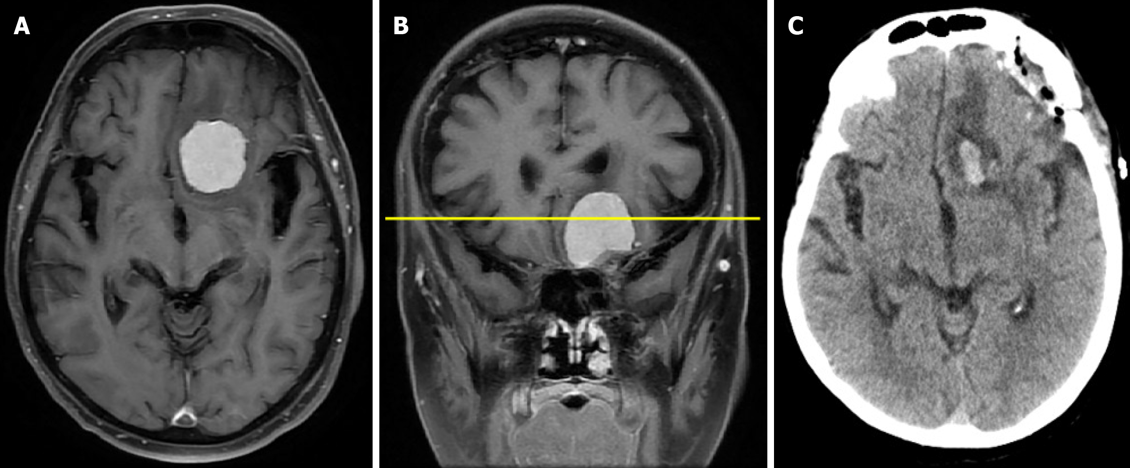
Figure 1 Cranial computed tomography and magnetic resonance imaging.
A: Contrast-enhanced axial T1-weighted magnetic resonance image demonstrating an anterior cranial fossa meningioma with mass effect on the left frontal lobe; B: Coronal magnetic resonance image confirming the frontobasal location of the lesion; C: Postoperative cranial computed tomography scan showing satisfactory postoperative findings.
- Citation: Begagić E, Bečulić H, Mašović A, Alić F, Huremović M, Vranić S. Carbamazepine-induced hyponatremia following meningioma surgery: A case report. World J Clin Cases 2026; 14(9): 118634
- URL: https://www.wjgnet.com/2307-8960/full/v14/i9/118634.htm
- DOI: https://dx.doi.org/10.12998/wjcc.v14.i9.118634