
















Copyright: ©Author(s) 2026.
World J Clin Cases. Mar 16, 2026; 14(8): 118789
Published online Mar 16, 2026. doi: 10.12998/wjcc.v14.i8.118789
Published online Mar 16, 2026. doi: 10.12998/wjcc.v14.i8.118789
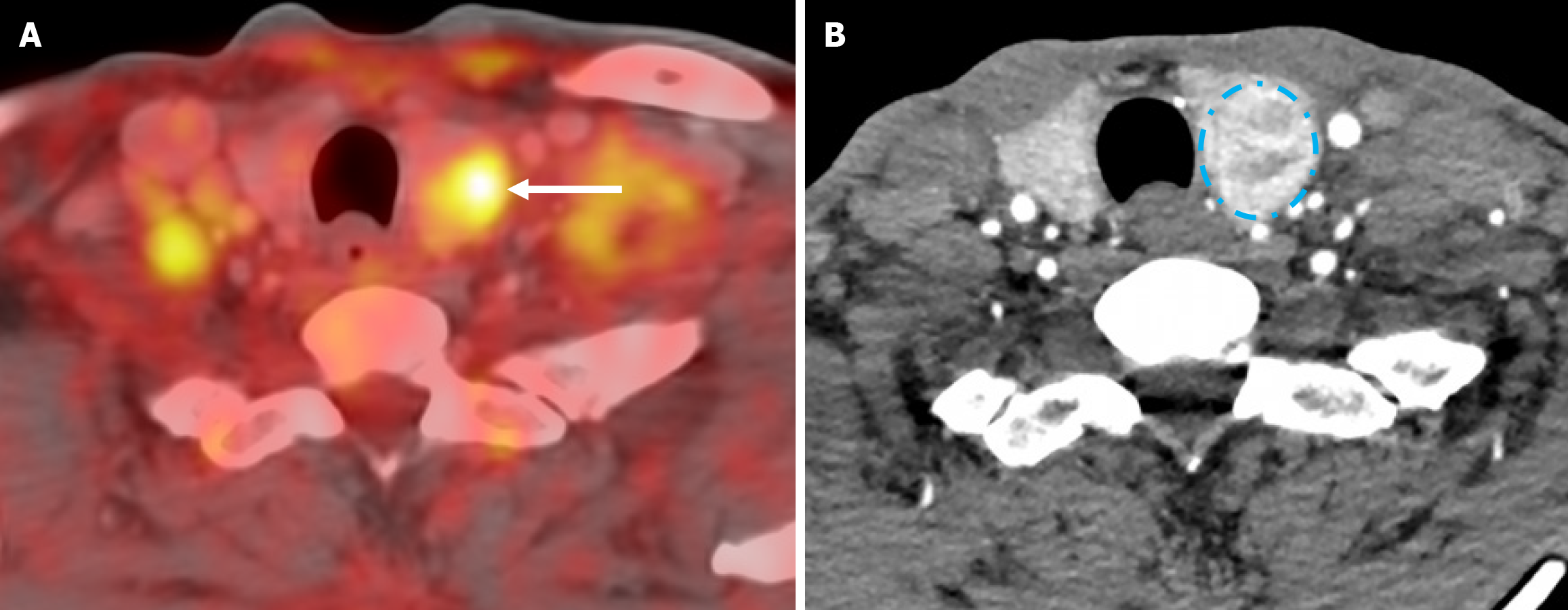
Figure 7 Positron emission tomography/computed tomography (level of the neck).
A: Axial view showing hypermetabolic left thyroid nodule (arrows); B: Contrast enhanced computed tomography neck axial showing heterogeneously enhancing left thyroid nodule.
- Citation: Paw LJ, Tan JK, Teoh CX, Megat Mohamad Aminuddin PNA, Agusalim AF, Cheong XK, Payus AO, Wan Nik Ahmad Mustafa WAK, Omar UA, Mustafa N, Chong GY, Tumian NR, Abdul Wahid SF, Chong CY, Tan HJ, Remli R. Castleman disease variant of polyneuropathy, organomegaly, endocrinopathy, monoclonal protein and skin changes syndrome: A case report. World J Clin Cases 2026; 14(8): 118789
- URL: https://www.wjgnet.com/2307-8960/full/v14/i8/118789.htm
- DOI: https://dx.doi.org/10.12998/wjcc.v14.i8.118789