
















Copyright: ©Author(s) 2026.
World J Clin Cases. Mar 16, 2026; 14(8): 118789
Published online Mar 16, 2026. doi: 10.12998/wjcc.v14.i8.118789
Published online Mar 16, 2026. doi: 10.12998/wjcc.v14.i8.118789
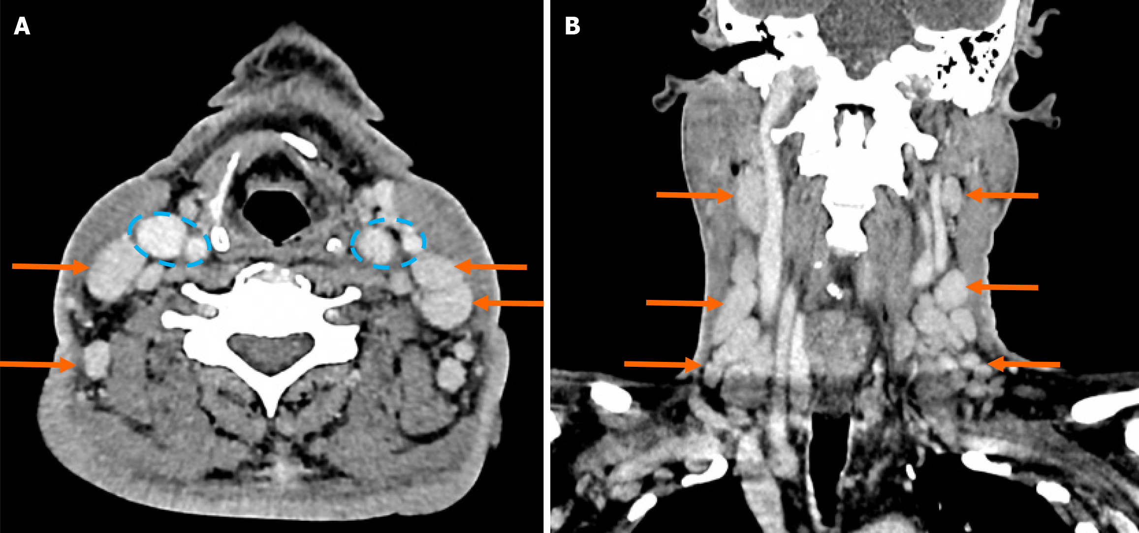
Figure 5 Contrast enhanced computed tomography neck.
A: Axial computed tomography (CT) with multiple enlarged cervical and supraclavicular lymph nodes (arrows) and patent bilateral carotid and jugular vessels are highlighted within the blue dotted lines; B: Sagittal CT with similarly enlarged nodes as mentioned (arrows).
- Citation: Paw LJ, Tan JK, Teoh CX, Megat Mohamad Aminuddin PNA, Agusalim AF, Cheong XK, Payus AO, Wan Nik Ahmad Mustafa WAK, Omar UA, Mustafa N, Chong GY, Tumian NR, Abdul Wahid SF, Chong CY, Tan HJ, Remli R. Castleman disease variant of polyneuropathy, organomegaly, endocrinopathy, monoclonal protein and skin changes syndrome: A case report. World J Clin Cases 2026; 14(8): 118789
- URL: https://www.wjgnet.com/2307-8960/full/v14/i8/118789.htm
- DOI: https://dx.doi.org/10.12998/wjcc.v14.i8.118789