
















Copyright: ©Author(s) 2026.
World J Clin Cases. Mar 16, 2026; 14(8): 117957
Published online Mar 16, 2026. doi: 10.12998/wjcc.v14.i8.117957
Published online Mar 16, 2026. doi: 10.12998/wjcc.v14.i8.117957
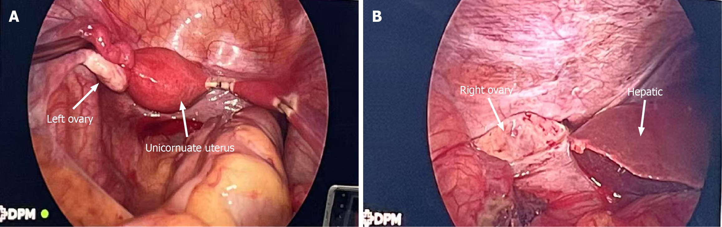
Figure 3 Intraoperative and histopathological correlation.
A: Laparoscopic view showing a left unicornuate uterus and normal left ovary within the pelvic cavity (arrow); B: Intraoperative photograph shows the ectopic fallopian tube and ovary (arrow) displaced to the right upper abdomen, firmly adherent to the inferior hepatic surface and capsule.
- Citation: Huang WJ, Zhao XR, Cao YQ, Huang XZ, Lai WL, Xi Y, Wu HY. Subhepatic ectopic pregnancy in Müllerian anomaly localized by large-field-of-view magnetic resonance imaging: A case report. World J Clin Cases 2026; 14(8): 117957
- URL: https://www.wjgnet.com/2307-8960/full/v14/i8/117957.htm
- DOI: https://dx.doi.org/10.12998/wjcc.v14.i8.117957