
















Copyright: ©Author(s) 2026.
World J Clin Cases. Mar 16, 2026; 14(8): 117957
Published online Mar 16, 2026. doi: 10.12998/wjcc.v14.i8.117957
Published online Mar 16, 2026. doi: 10.12998/wjcc.v14.i8.117957
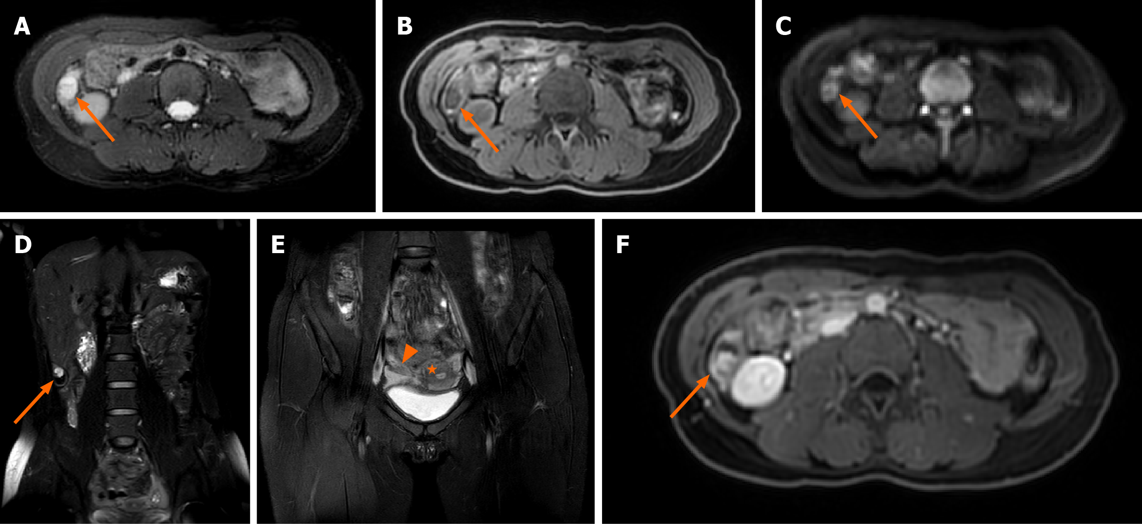
Figure 1 Magnetic resonance imaging of a 28-year-old pregnant woman with surgically and pathologically confirmed unruptured tubal ectopic pregnancy.
A: Axial T2-weighted fat-suppressed image shows a small nodular hyperintense focus (arrow) within the gestational sac; B: Axial T1-weighted image demonstrates the nodule (arrow) with isointense signal relative to muscle; C: Diffusion-weighted image (b-value = 800 s/mm²) shows no restricted diffusion within the lesion (arrow); D: The coronal magnetic resonance image of the entire abdomen shows that the gestational sac (arrow) is located between the lower edge of the right lobe of the liver and the right iliac fossa; E: Coronal T2-weighted image confirms uterine morphology consistent with a left unicornuate uterus (star) and a right rudimentary horn (triangle). The uterus is deviated toward the left pelvic sidewall; F: Axial contrast-enhanced T1-weighted fat-suppressed image reveals vivid enhancement of the nodule (arrow) and mild enhancement of the cyst wall.
- Citation: Huang WJ, Zhao XR, Cao YQ, Huang XZ, Lai WL, Xi Y, Wu HY. Subhepatic ectopic pregnancy in Müllerian anomaly localized by large-field-of-view magnetic resonance imaging: A case report. World J Clin Cases 2026; 14(8): 117957
- URL: https://www.wjgnet.com/2307-8960/full/v14/i8/117957.htm
- DOI: https://dx.doi.org/10.12998/wjcc.v14.i8.117957