
















©Author(s) (or their employer(s)) 2026.
World J Clin Cases. Mar 6, 2026; 14(7): 116694
Published online Mar 6, 2026. doi: 10.12998/wjcc.v14.i7.116694
Published online Mar 6, 2026. doi: 10.12998/wjcc.v14.i7.116694
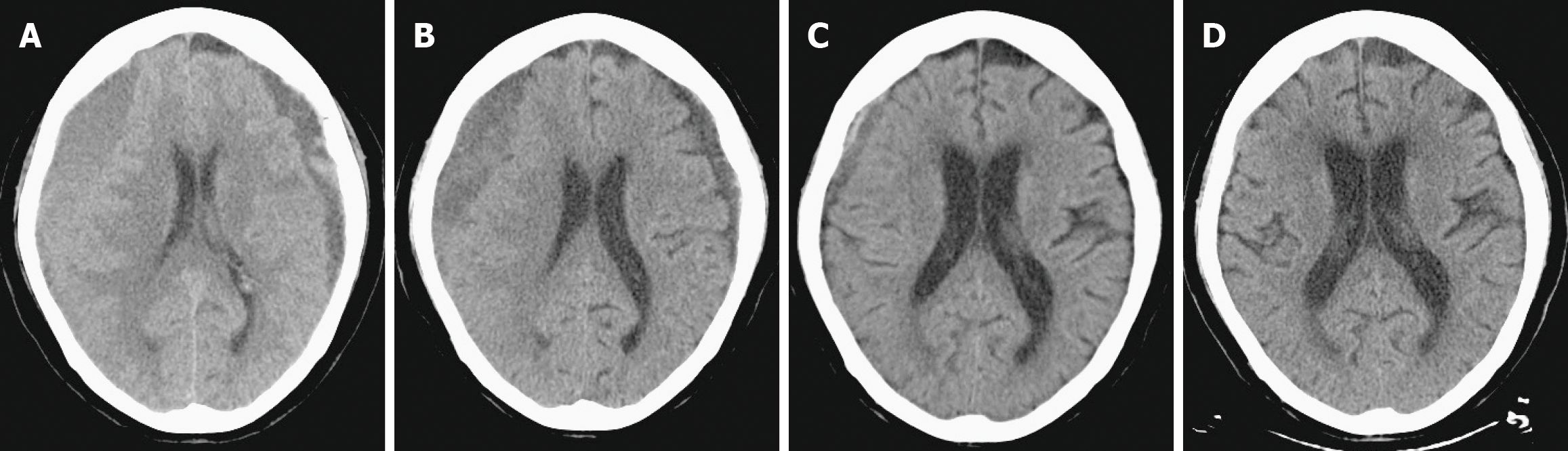
Figure 3 Axial brain computed tomography showing progressive resolution of chronic subdural hematoma following middle meningeal artery embolization.
A: Pre-embolization image illustrating extensive bilateral chronic subdural hematoma; B: One-month post-embolization image showing substantial hematoma reduction; C: Two-month post-embolization image demonstrating continued hematoma clearance; D: Three-month post-embolization image showing complete hematoma resolution and excellent brain re-expansion.
- Citation: Chen CY, Hsu YH. Complete resolution of massive chronic subdural hematoma with refined layered middle meningeal artery embolization: A case report. World J Clin Cases 2026; 14(7): 116694
- URL: https://www.wjgnet.com/2307-8960/full/v14/i7/116694.htm
- DOI: https://dx.doi.org/10.12998/wjcc.v14.i7.116694