
















©The Author(s) 2026.
World J Clin Cases. Feb 26, 2026; 14(6): 118545
Published online Feb 26, 2026. doi: 10.12998/wjcc.v14.i6.118545
Published online Feb 26, 2026. doi: 10.12998/wjcc.v14.i6.118545
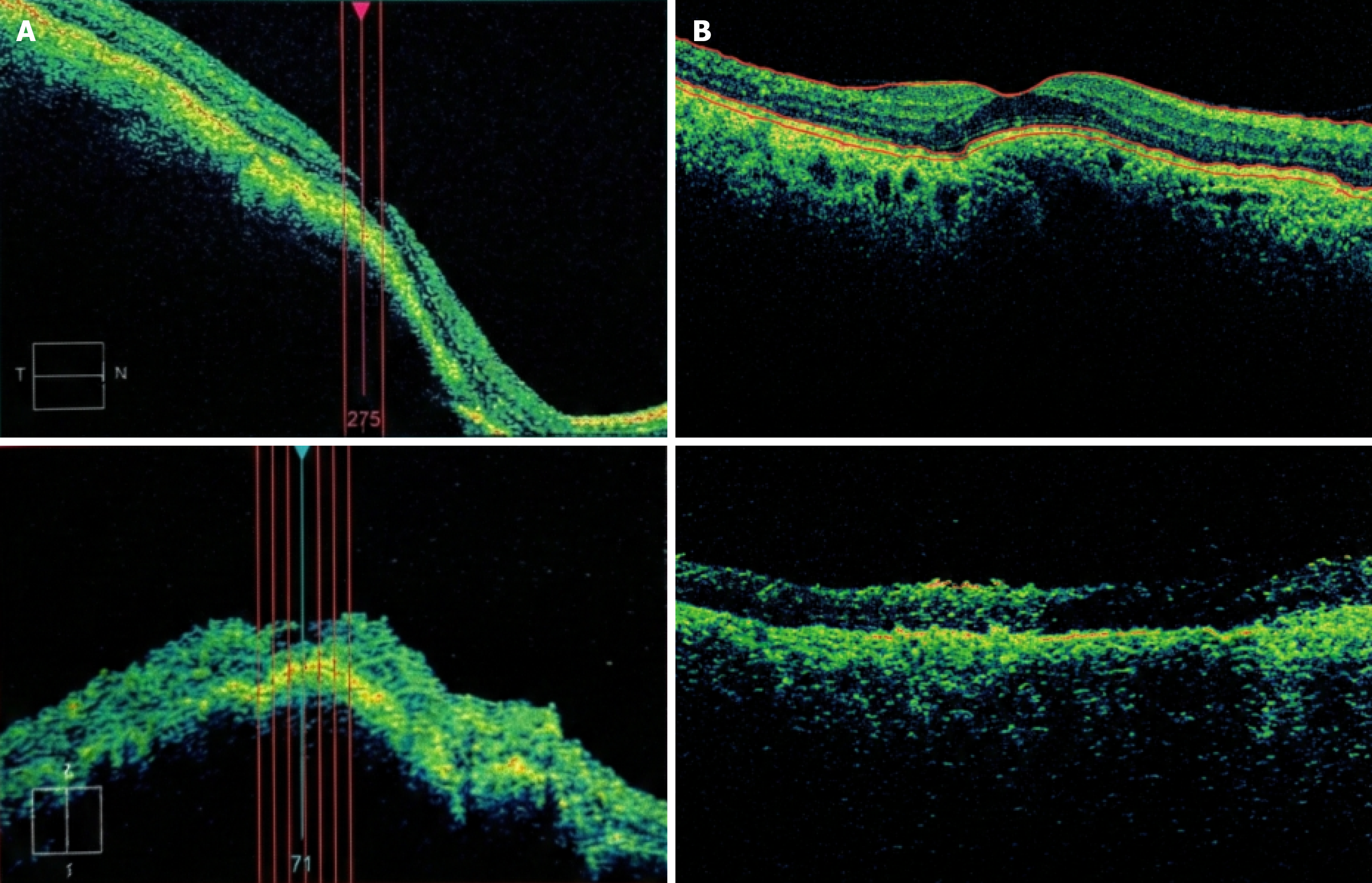
Figure 1 Case 1 optical coherence tomography (baseline and follow-up).
A: Baseline (preoperative) spectral-domain optical coherence tomography (OCT) of the right eye demonstrating fovea-involving submacular hemorrhage secondary to choroidal neovascularization; B: Postoperative follow-up OCT (approximately 4 weeks) demonstrating interval postoperative evolution compared with baseline following pars plana vitrectomy with subretinal tissue plasminogen activator and gas tamponade. Representative horizontal B-scan through the fovea is shown.
- Citation: El Mollayess G, Jaroudi M, Tlaiss Y, Itaoui R, Abiad B. Surgical management of acute choroidal neovascularization related submacular hemorrhage: Three case reports. World J Clin Cases 2026; 14(6): 118545
- URL: https://www.wjgnet.com/2307-8960/full/v14/i6/118545.htm
- DOI: https://dx.doi.org/10.12998/wjcc.v14.i6.118545