















©The Author(s) 2026.
World J Clin Cases. Feb 26, 2026; 14(6): 118184
Published online Feb 26, 2026. doi: 10.12998/wjcc.v14.i6.118184
Published online Feb 26, 2026. doi: 10.12998/wjcc.v14.i6.118184
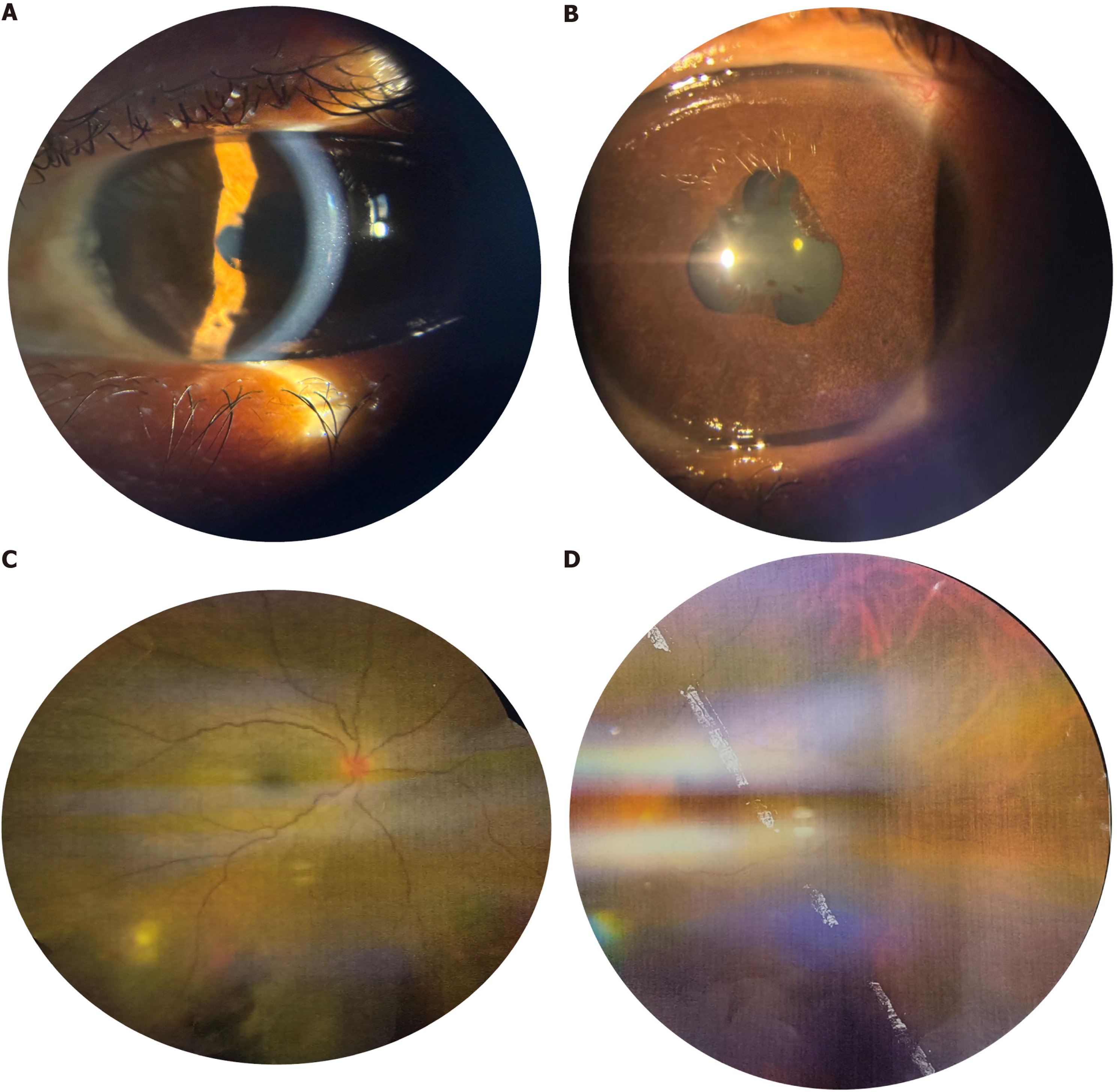
Figure 1 Clinical ocular manifestations of chronic panuveitis.
A: Slit-lamp photograph of the left eye demonstrating diffuse, flat, pigmented keratic preci
- Citation: Singh N, Morya AK, Walia S, Udenia H. Tubercular chorioretinitis mimicking sarcoidosis in a patient with celiac disease and erythema nodosum: A case report. World J Clin Cases 2026; 14(6): 118184
- URL: https://www.wjgnet.com/2307-8960/full/v14/i6/118184.htm
- DOI: https://dx.doi.org/10.12998/wjcc.v14.i6.118184