
















©The Author(s) 2026.
World J Clin Cases. Feb 26, 2026; 14(6): 118135
Published online Feb 26, 2026. doi: 10.12998/wjcc.v14.i6.118135
Published online Feb 26, 2026. doi: 10.12998/wjcc.v14.i6.118135
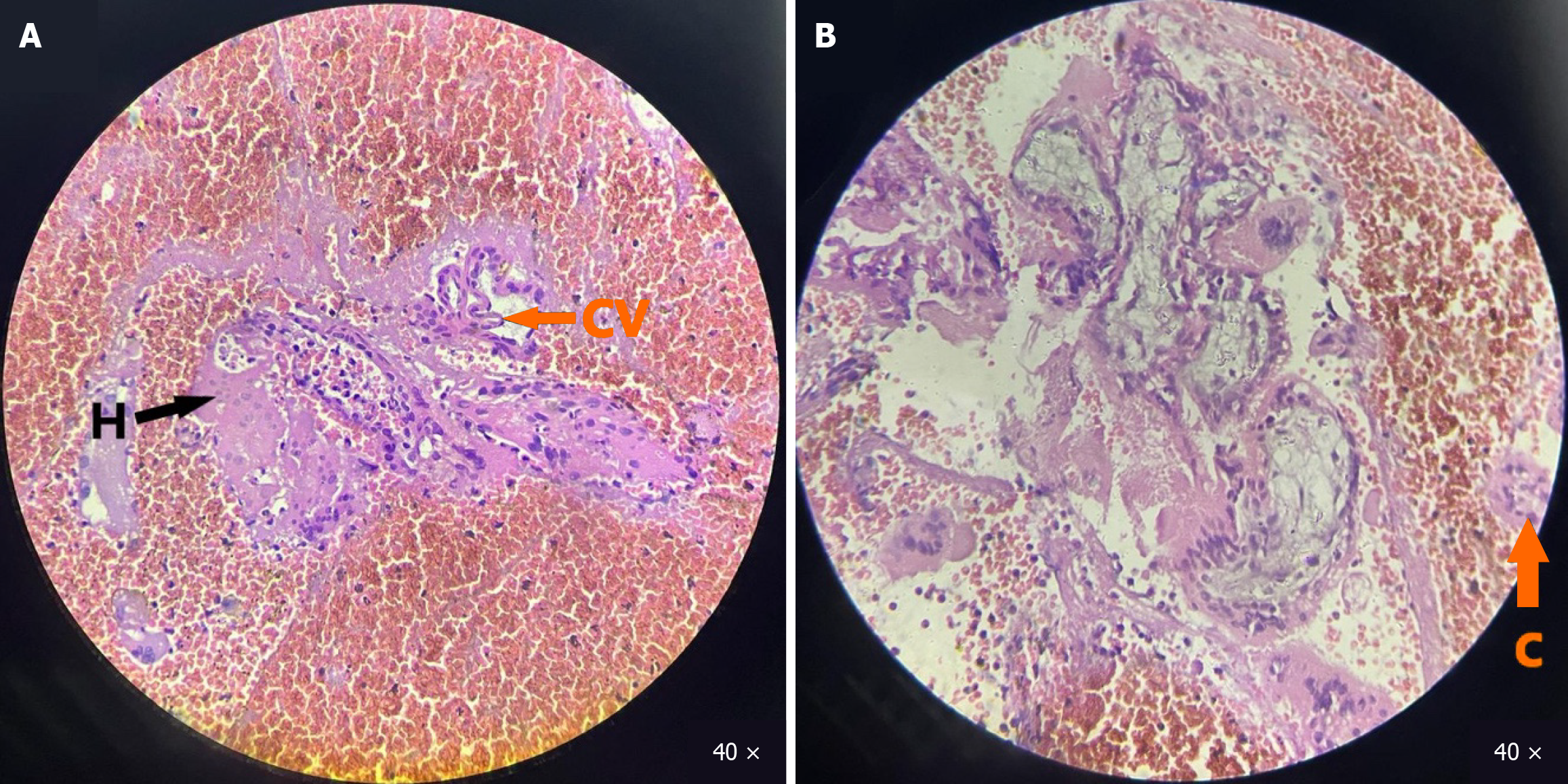
Figure 5 Histopathological examination.
A: Magnification (× 40) magnification of hepatocytes (black arrow) and chorionic villi (orange arrow) with peripheral area of necrosis and hemorrhage; B: Cytotrophoblasts visualized (orange arrow) along with inflammatory infiltrates. S: Synctiotrophoblasts; H: Hepatocytes; CV: Chorionic villi; C: Cytotrophoblasts.
- Citation: Bhati G, Mongardini FM, Bhati K, Singh P, Bansal R, Bansal A, Mahajan S, Docimo L, Caricato M, Capolupo GT, Carannante F. Ruptured primary intrahepatic ectopic pregnancy: A case report and review of literature. World J Clin Cases 2026; 14(6): 118135
- URL: https://www.wjgnet.com/2307-8960/full/v14/i6/118135.htm
- DOI: https://dx.doi.org/10.12998/wjcc.v14.i6.118135