
















©The Author(s) 2026.
World J Clin Cases. Feb 26, 2026; 14(6): 118135
Published online Feb 26, 2026. doi: 10.12998/wjcc.v14.i6.118135
Published online Feb 26, 2026. doi: 10.12998/wjcc.v14.i6.118135
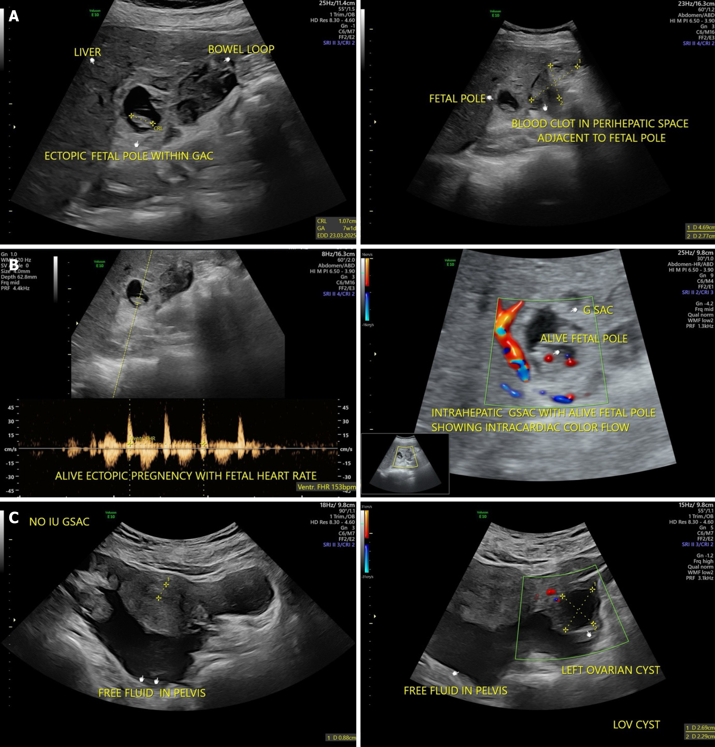
Figure 1 Abdominal ultrasound.
A: Ultrasound imaging demonstrated a large volume of free intraperitoneal fluid that was consistent with massive hemoperitoneum; B: Live ectopic pregnancy with positive fetal heart rate. Doppler study showed intracardiac color flow; C: Focused abdominal ultrasonography of the liver. Ultrasound imaging showed a heterogeneous lesion with mixed echogenicity arising from the right hepatic lobe that was suspicious of ectopic gestational implantation.
- Citation: Bhati G, Mongardini FM, Bhati K, Singh P, Bansal R, Bansal A, Mahajan S, Docimo L, Caricato M, Capolupo GT, Carannante F. Ruptured primary intrahepatic ectopic pregnancy: A case report and review of literature. World J Clin Cases 2026; 14(6): 118135
- URL: https://www.wjgnet.com/2307-8960/full/v14/i6/118135.htm
- DOI: https://dx.doi.org/10.12998/wjcc.v14.i6.118135