
















©The Author(s) 2026.
World J Clin Cases. Feb 26, 2026; 14(6): 117655
Published online Feb 26, 2026. doi: 10.12998/wjcc.v14.i6.117655
Published online Feb 26, 2026. doi: 10.12998/wjcc.v14.i6.117655
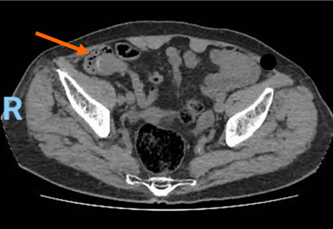
Figure 3 Computed tomography scan with intravenous contrast showing a well-circumscribed, low fluid attenuation tubular-shaped structure, seen in continuation with the base of the cecum, grossly measuring 64 mm × 29 mm × 28 mm, corresponding to the appendix (orange arrow).
- Citation: Aby Hadeer R, Ghattas S, Farhat H, Maalouf H, Bitar JE, Ayash D, Mohtar F, Elias B, Wakim R. Cytoreductive surgery and hyperthermic intraperitoneal chemotherapy for appendiceal mucocele tumors: Five case reports and review of literature. World J Clin Cases 2026; 14(6): 117655
- URL: https://www.wjgnet.com/2307-8960/full/v14/i6/117655.htm
- DOI: https://dx.doi.org/10.12998/wjcc.v14.i6.117655