
















©The Author(s) 2026.
World J Clin Cases. Feb 6, 2026; 14(4): 117314
Published online Feb 6, 2026. doi: 10.12998/wjcc.v14.i4.117314
Published online Feb 6, 2026. doi: 10.12998/wjcc.v14.i4.117314
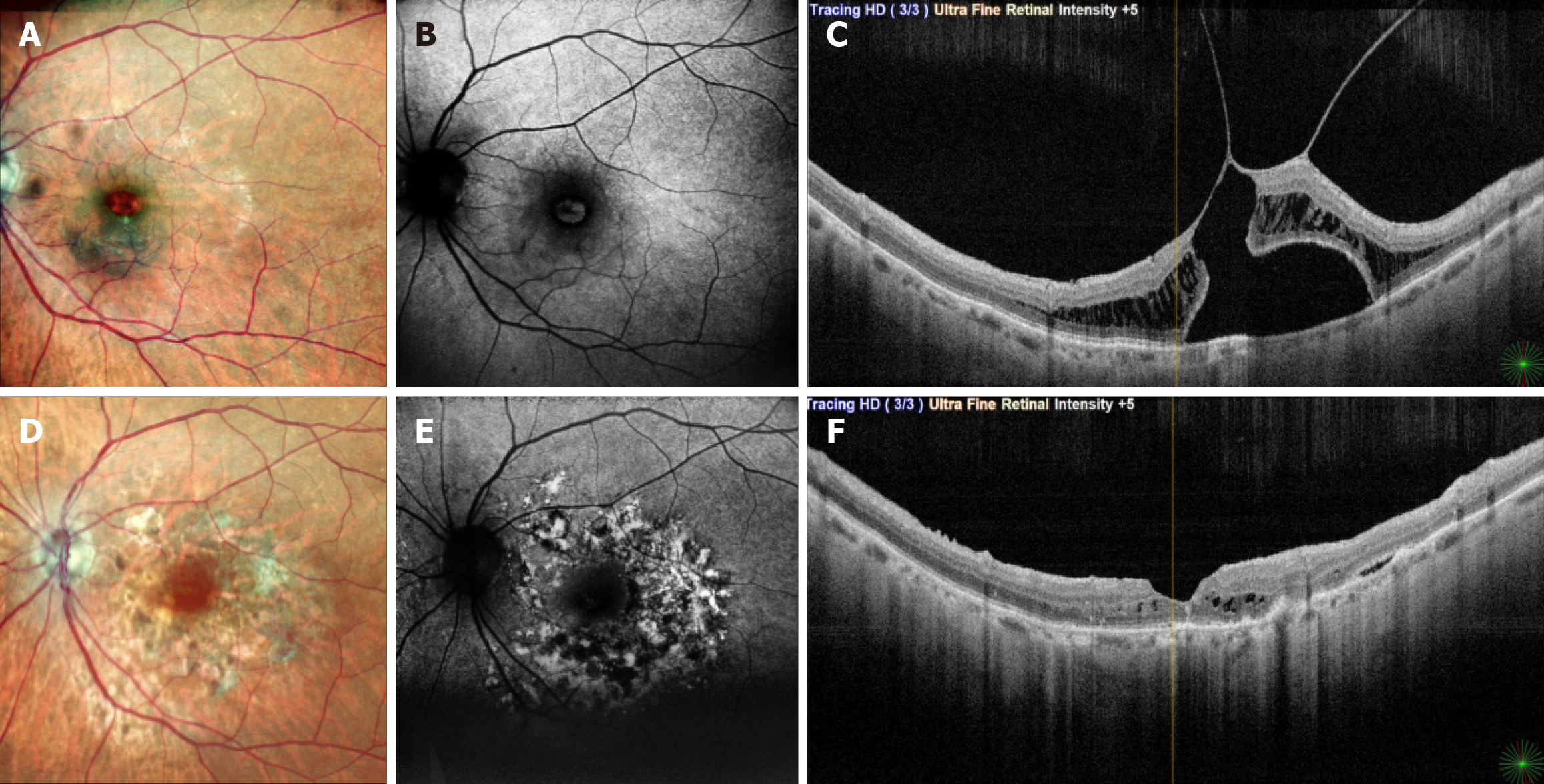
Figure 3 Imaging features before and after surgery – case 9.
A-C: Preoperative imaging of patient 9, who underwent surgery for vitreomacular traction (VMT) with foveal detachment in the left eye. Initial retinal thickness measured 468 µm, and visual acuity was 6/18. Preoperative optical coherence tomography (OCT) shows significant VMT with neurosensory detachment involving the fovea, intact outer retinal layers, intact retinal pigment epithelium (RPE), and a disrupted external limiting membrane. Fundus autofluorescence imaging of the left eye normal autofluorescence signal patterns at the fovea. Internal limiting membrane (ILM) staining was performed under balanced salt solution for 2 minutes, with a total surgical time of 2 hours. One-month postoperative OCT showing brilliant blue G (BBG)-related retinal toxicity sparing the fovea; D-F: At 5 months post-surgery, the foveal contour was restored with visual acuity improvement to 6/9. Fundus autofluorescence identified abnormal hyper- and hypoautofluorescent signals indicative of BBG-related retinal toxicity. OCT at 5 months demonstrated outer retinal damage and RPE mottling, sparing the fovea. The parafoveal damage corresponded to the ILM peel area.
- Citation: Venkatesh R, James E, Hande P, Raj P, Tendulkar K, Prabhu V. Retinal architecture as a determinant of brilliant blue G phototoxicity during internal limiting membrane peeling: A case series. World J Clin Cases 2026; 14(4): 117314
- URL: https://www.wjgnet.com/2307-8960/full/v14/i4/117314.htm
- DOI: https://dx.doi.org/10.12998/wjcc.v14.i4.117314