
















©The Author(s) 2026.
World J Clin Cases. Feb 6, 2026; 14(4): 117314
Published online Feb 6, 2026. doi: 10.12998/wjcc.v14.i4.117314
Published online Feb 6, 2026. doi: 10.12998/wjcc.v14.i4.117314
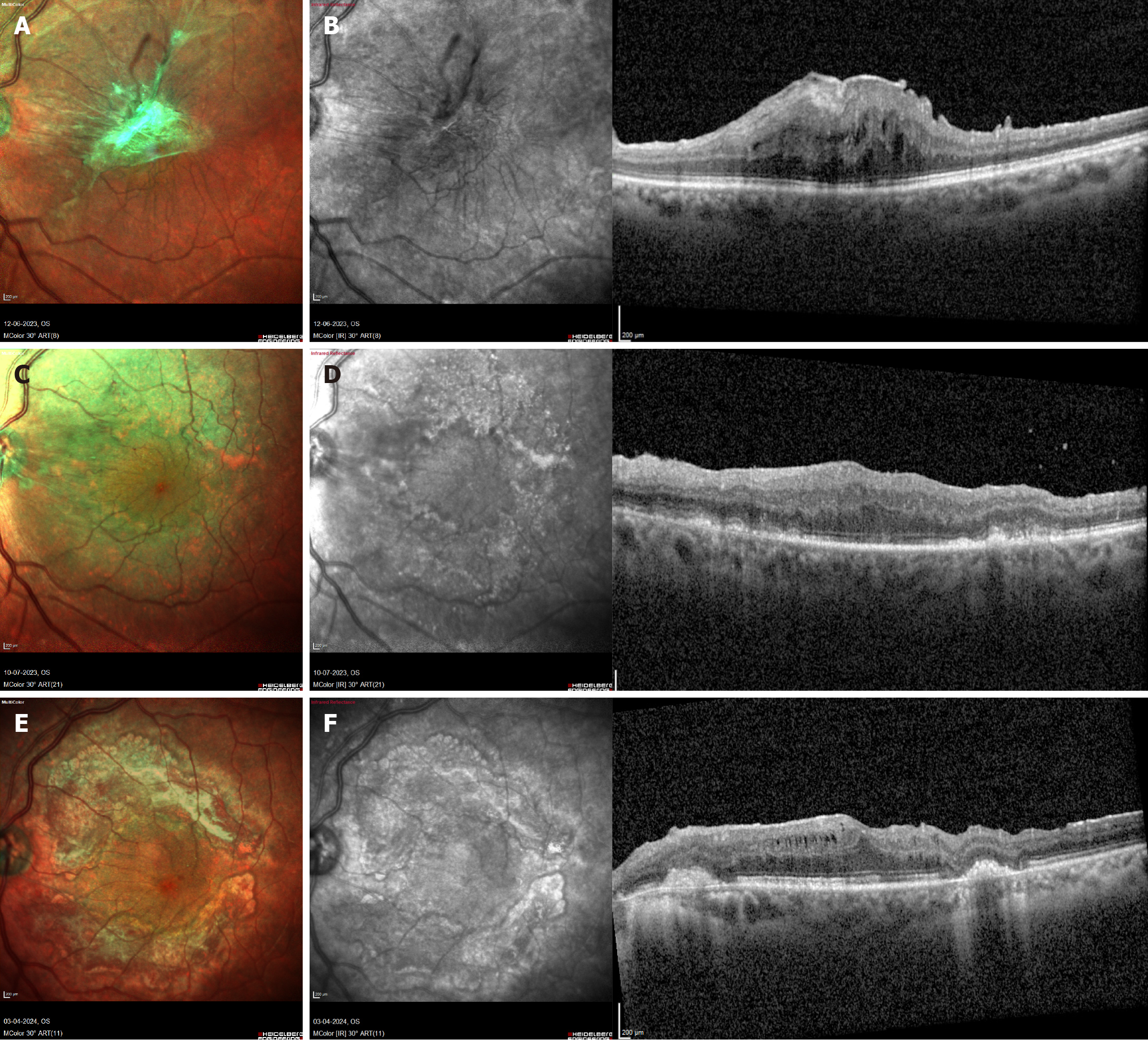
Figure 2 Imaging features before and after surgery - case 8.
A and B: Correspond to patient 8, who underwent surgery for an epiretinal membrane in the left eye, presenting with retinal traction and increased retinal thickness (590 µm). The initial visual acuity was 6/30. Preoperative optical coherence tomography (OCT) showed significant retinal traction caused by the thick epiretinal membrane, with intact outer retinal layers and retinal pigment epithelium (RPE) at the fovea. ILM staining was performed twice under air for a total of 3.5 minutes, with a surgical time of 110 minutes; C and D: Ten days postoperatively, OCT revealed outer retinal layer damage and RPE mottling, sparing the fovea. The parafoveal damage corresponded to the ILM peel area, with visual acuity improving to 6/24; E and F: At 10 months post-surgery, further reduction in foveal thickness was observed, with visual acuity improving to 6/8 and the brilliant blue G related retinal toxicity sparing the fovea.
- Citation: Venkatesh R, James E, Hande P, Raj P, Tendulkar K, Prabhu V. Retinal architecture as a determinant of brilliant blue G phototoxicity during internal limiting membrane peeling: A case series. World J Clin Cases 2026; 14(4): 117314
- URL: https://www.wjgnet.com/2307-8960/full/v14/i4/117314.htm
- DOI: https://dx.doi.org/10.12998/wjcc.v14.i4.117314