
















©The Author(s) 2026.
World J Clin Cases. Feb 6, 2026; 14(4): 117226
Published online Feb 6, 2026. doi: 10.12998/wjcc.v14.i4.117226
Published online Feb 6, 2026. doi: 10.12998/wjcc.v14.i4.117226
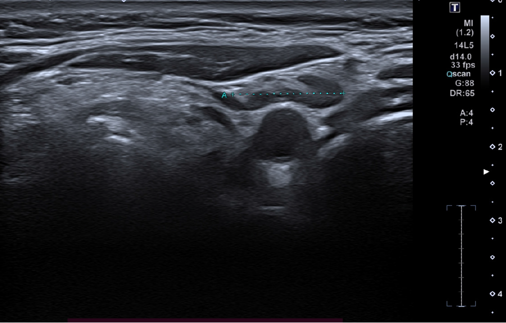
Figure 2 High-frequency ultrasound (24 MHz) image of the left laterocervical region showing a single lymph node (outlined in blue dashed line), not suspicious for metastatic involvement.
The node presents with preserved morphology, including a visible fatty hilum and a well-defined cortex, with no signs of pathological enlargement or altered echogenicity.
- Citation: Russo A, Patanè V, Pezzella MC, Troiani T, Argenziano G, Reginelli A. Ultra-high-frequency ultrasound in the detection of recurrent conjunctival melanoma: A case report. World J Clin Cases 2026; 14(4): 117226
- URL: https://www.wjgnet.com/2307-8960/full/v14/i4/117226.htm
- DOI: https://dx.doi.org/10.12998/wjcc.v14.i4.117226