
















©The Author(s) 2026.
World J Clin Cases. Feb 6, 2026; 14(4): 116737
Published online Feb 6, 2026. doi: 10.12998/wjcc.v14.i4.116737
Published online Feb 6, 2026. doi: 10.12998/wjcc.v14.i4.116737
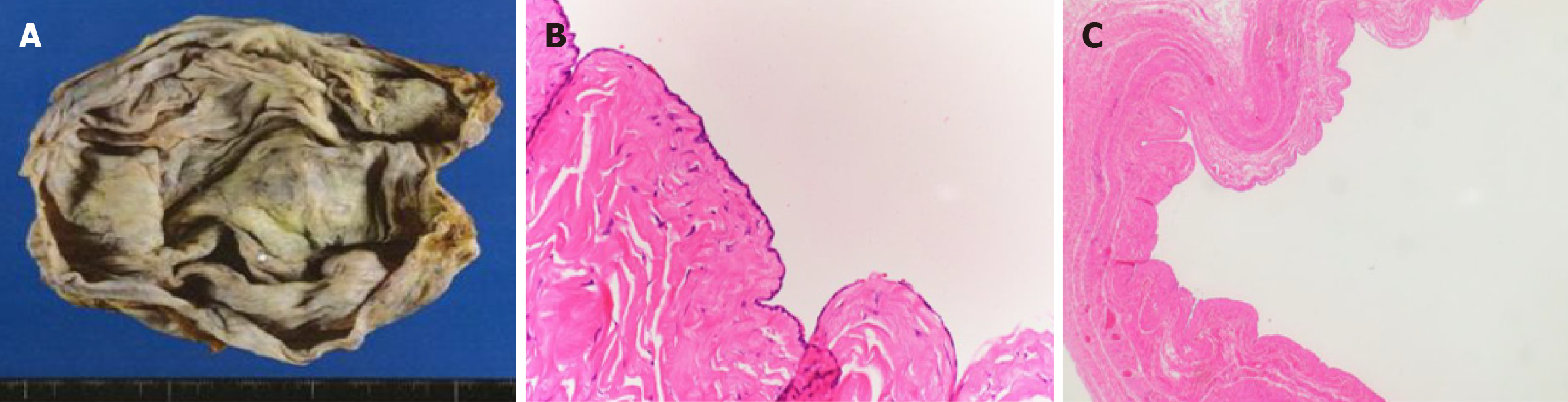
Figure 3 Resected specimen and histopathological findings.
A: The inner surface of the cyst was smooth, with no evidence of solid proliferation; B and C: The cystic inner surface was lined by cuboidal epithelium without atypia, findings suggestive of cystadenoma. No features suspicious for malignancy were observed. Hematoxylin and eosin staining; B: × 20, C: × 400.
- Citation: Segawa A, Kakinuma T, Miyazawa C, Takeuchi J, Tamura M, Morita A, Takae S, Suzuki N, Ariizumi Y. Successful perioperative management for a giant ovarian tumor in older adults: A case report. World J Clin Cases 2026; 14(4): 116737
- URL: https://www.wjgnet.com/2307-8960/full/v14/i4/116737.htm
- DOI: https://dx.doi.org/10.12998/wjcc.v14.i4.116737