
















©The Author(s) 2026.
World J Clin Cases. Feb 6, 2026; 14(4): 116648
Published online Feb 6, 2026. doi: 10.12998/wjcc.v14.i4.116648
Published online Feb 6, 2026. doi: 10.12998/wjcc.v14.i4.116648
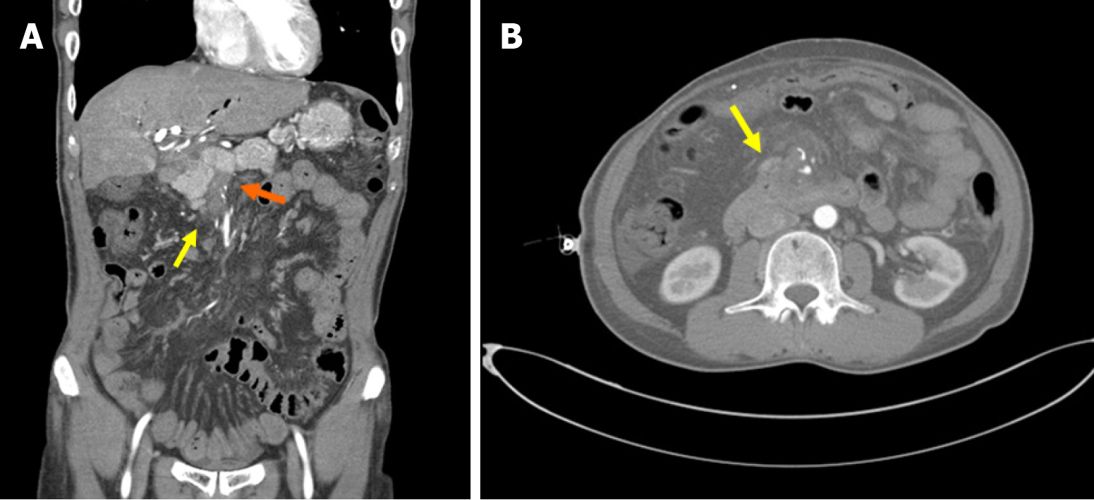
Figure 2 Abdominal computed tomography.
A: On the coronal view, there was portal vein (orange arrow) and superior mesenteric vein thrombus as well as duodenal varix of 3rd portion (yellow arrow); B: On the axial view, there was duodenal varix at the 3rd portion (yellow arrow).
- Citation: Lee H, Han YH, Chung JW, Kim KO, Kwon KA, Kim JH. Upper gastrointestinal bleeding with duodenal varix: A case report. World J Clin Cases 2026; 14(4): 116648
- URL: https://www.wjgnet.com/2307-8960/full/v14/i4/116648.htm
- DOI: https://dx.doi.org/10.12998/wjcc.v14.i4.116648