
















Copyright: ©Author(s) 2026.
World J Clin Cases. Oct 26, 2026; 14(28): 127786
Published online Oct 26, 2026. doi: 10.12998/wjcc.127786
Published online Oct 26, 2026. doi: 10.12998/wjcc.127786
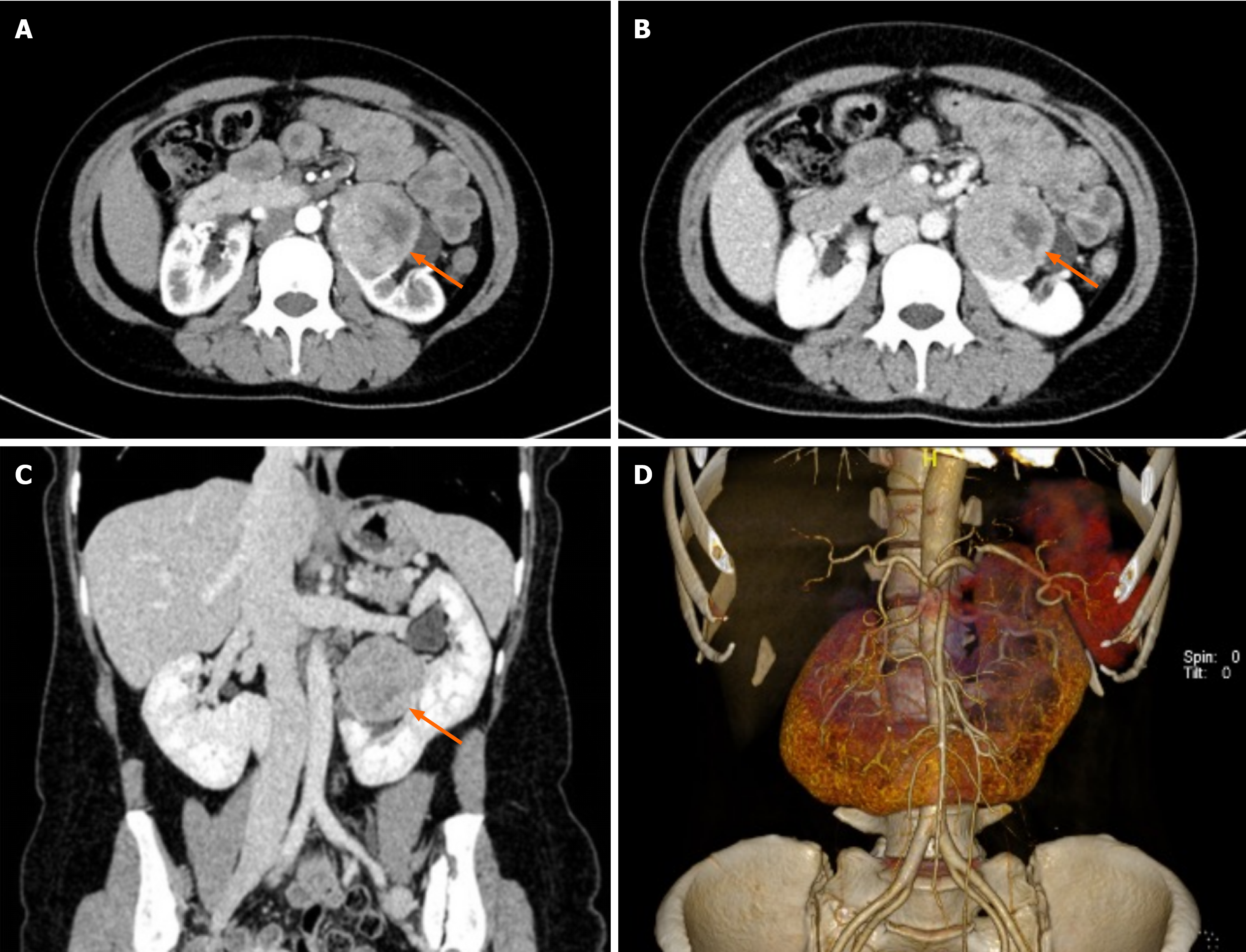
Figure 1 Computed tomography urography findings of primary renal carcinoid tumor within horseshoe kidney.
A: Axial contrast-enhanced computed tomography (CT) image (nephrographic phase): Demonstrates a heterogeneous-density mass (orange arrow) within the left-sided moiety of the horseshoe kidney, with a central low-density necrotic area visible; B: Another axial CT slice shows mass boundary relative to fused renal parenchyma (orange arrow); C: Coronal reformatted computed tomography urography image shows tumor location adjacent to horseshoe-kidney renal isthmus (orange arrow); D: Three-dimensional volume-rendering reconstruction demonstrates horseshoe-kidney fused lower-pole anatomical configuration and spatial relationship of the mass.
- Citation: Zhao Y, Wu XC. Primary renal carcinoid tumor in a horseshoe kidney presenting with constipation: A case report. World J Clin Cases 2026; 14(28): 127786
- URL: https://www.wjgnet.com/2307-8960/full/v14/i28/127786.htm
- DOI: https://dx.doi.org/10.12998/wjcc.127786