
















Copyright: ©Author(s) 2026.
World J Clin Cases. Oct 26, 2026; 14(28): 127546
Published online Oct 26, 2026. doi: 10.12998/wjcc.127546
Published online Oct 26, 2026. doi: 10.12998/wjcc.127546
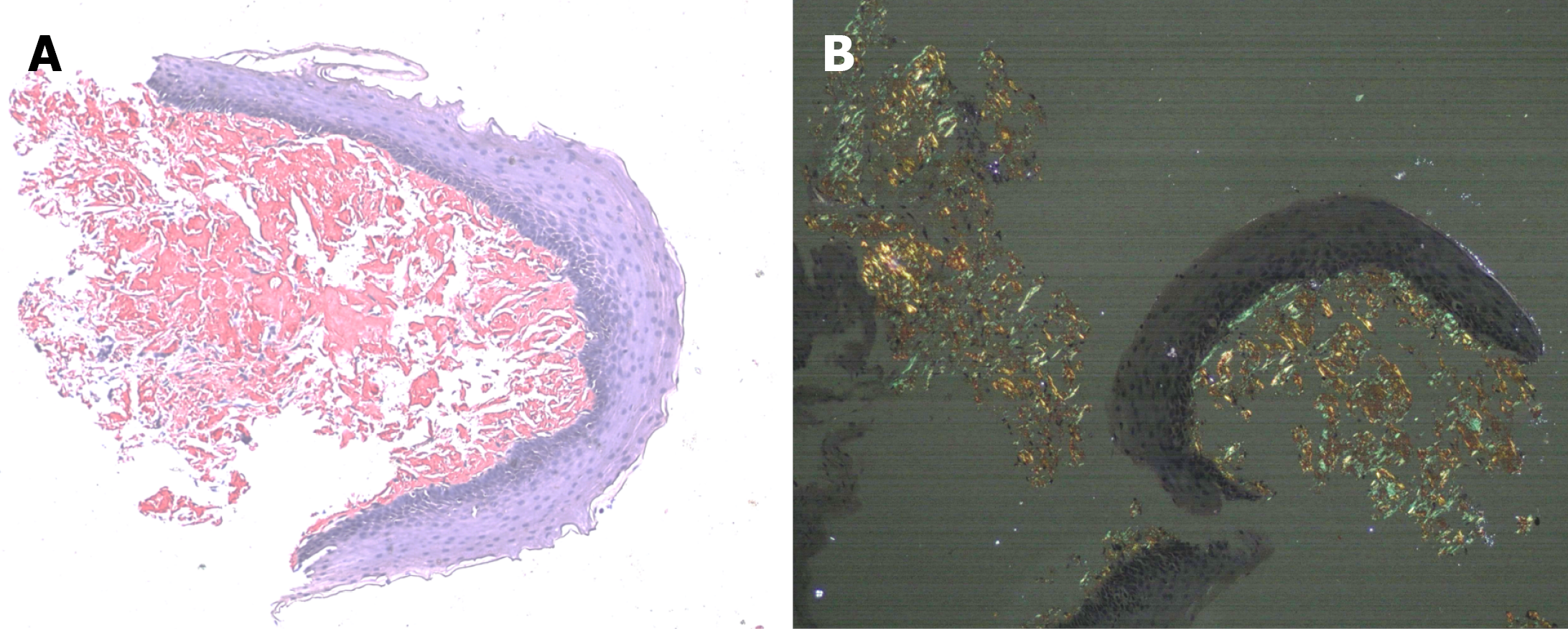
Figure 3 Congo red staining confirming amyloid deposition in the esophageal biopsy.
A: Congo red staining demonstrating salmon-pink amyloid deposits within the subepithelial stroma (×40); B: The same deposits showing characteristic apple-green birefringence under polarized light microscopy, confirming amyloid deposition (×40).
- Citation: Sachdeva S, Sirohi N, Mandal K, Bansal D, Vaithiyam V, Dalal A, Goyal S, Sakhuja P. Esophageal stricture due to amyloidosis in plasma cell dyscrasia: A case report and review of literature. World J Clin Cases 2026; 14(28): 127546
- URL: https://www.wjgnet.com/2307-8960/full/v14/i28/127546.htm
- DOI: https://dx.doi.org/10.12998/wjcc.127546