
















Copyright: ©Author(s) 2026.
World J Clin Cases. Sep 26, 2026; 14(27): 126787
Published online Sep 26, 2026. doi: 10.12998/wjcc.126787
Published online Sep 26, 2026. doi: 10.12998/wjcc.126787
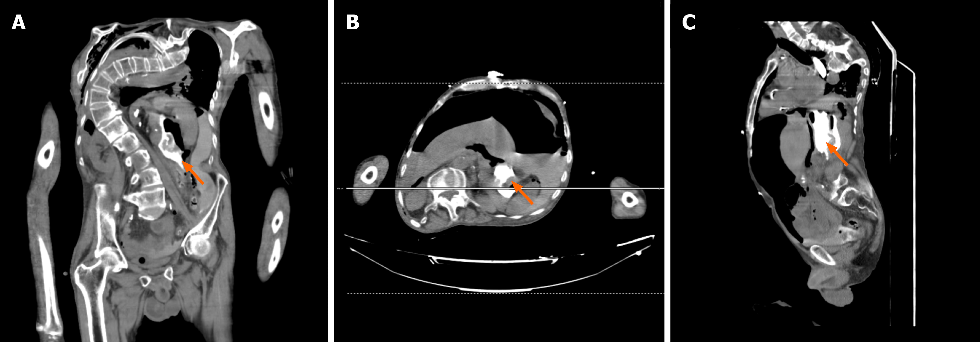
Figure 1 Contrast-enhanced computed tomography at emergency admission.
A: Coronal image showing extravasation of orally administered contrast material from the posterior wall of the gastric corpus (arrow), with free intraperitoneal fluid and marked thoracolumbar scoliosis; B: Axial image at the level of the gastric corpus demonstrating the posterior gastric wall defect with contrast extravasation (arrow), together with perigastric free air and fluid; C: Sagittal reconstruction delineating the posterior gastric wall defect with contrast leakage into the retrogastric space (arrow) and diffuse free fluid throughout the abdomen. All scanner overlays containing the examination date and accession number have been removed, and the images are fully de-identified.
- Citation: Cakiroglu OF, Gurbuz J, Taslidere B, Cander B. Posterior gastric wall perforation and acute kidney injury after upper endoscopy with esophageal dilation: A case report. World J Clin Cases 2026; 14(27): 126787
- URL: https://www.wjgnet.com/2307-8960/full/v14/i27/126787.htm
- DOI: https://dx.doi.org/10.12998/wjcc.126787