
















Copyright: ©Author(s) 2026.
World J Clin Cases. Sep 16, 2026; 14(26): 126834
Published online Sep 16, 2026. doi: 10.12998/wjcc.126834
Published online Sep 16, 2026. doi: 10.12998/wjcc.126834
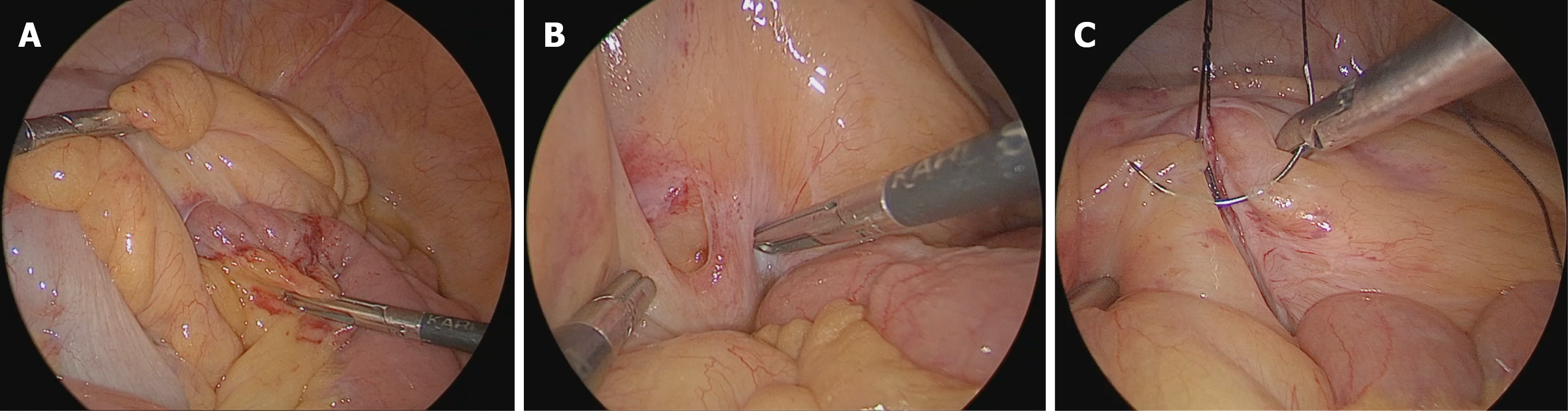
Figure 2 Laparoscopic view.
A: Intraoperative laparoscopic view at diagnostic laparoscopy. Small bowel loops are herniating through a defect in the transverse mesocolon. The herniated jejunal segment (center) shows minimal serosal congestion and a few petechial hemorrhages at the mesocolic defect, but remains pink, well perfused, and clearly viable, without edema or ischemic discoloration. Atraumatic graspers are used to retract the surrounding loops and delineate the defect margins before reduction; B: Laparoscopic view of the transverse mesocolic defect after reduction of the herniated bowel. An oval defect in the transverse mesocolon is seen (left of center) with intact, mildly hyperemic margins and no active bleeding. Healthy small bowel loops lie below. The reduced jejunum is pink and viable. A laparoscopic needle holder is positioned at the defect margin in preparation for primary closure with interrupted 2-0 silk sutures; C: Laparoscopic closure of the transverse mesocolic defect with interrupted 2-0 braided silk sutures. The edges of the mesocolic defect have been approximated with interrupted sutures, the final suture being passed with a curved needle held in the laparoscopic needle holder (right of center). The repaired mesocolic leaf is intact, with its vessels preserved and no bleeding at the suture line. The reduced jejunal loops lying below are uniformly pink and well perfused.
- Citation: Joshi M, Jha S, Jha S, Jhawar N. Laparoscopic treatment of a transmesocolic internal hernia causing small bowel obstruction: A case report. World J Clin Cases 2026; 14(26): 126834
- URL: https://www.wjgnet.com/2307-8960/full/v14/i26/126834.htm
- DOI: https://dx.doi.org/10.12998/wjcc.126834