
















Copyright: ©Author(s) 2026.
World J Clin Cases. Sep 16, 2026; 14(26): 126834
Published online Sep 16, 2026. doi: 10.12998/wjcc.126834
Published online Sep 16, 2026. doi: 10.12998/wjcc.126834
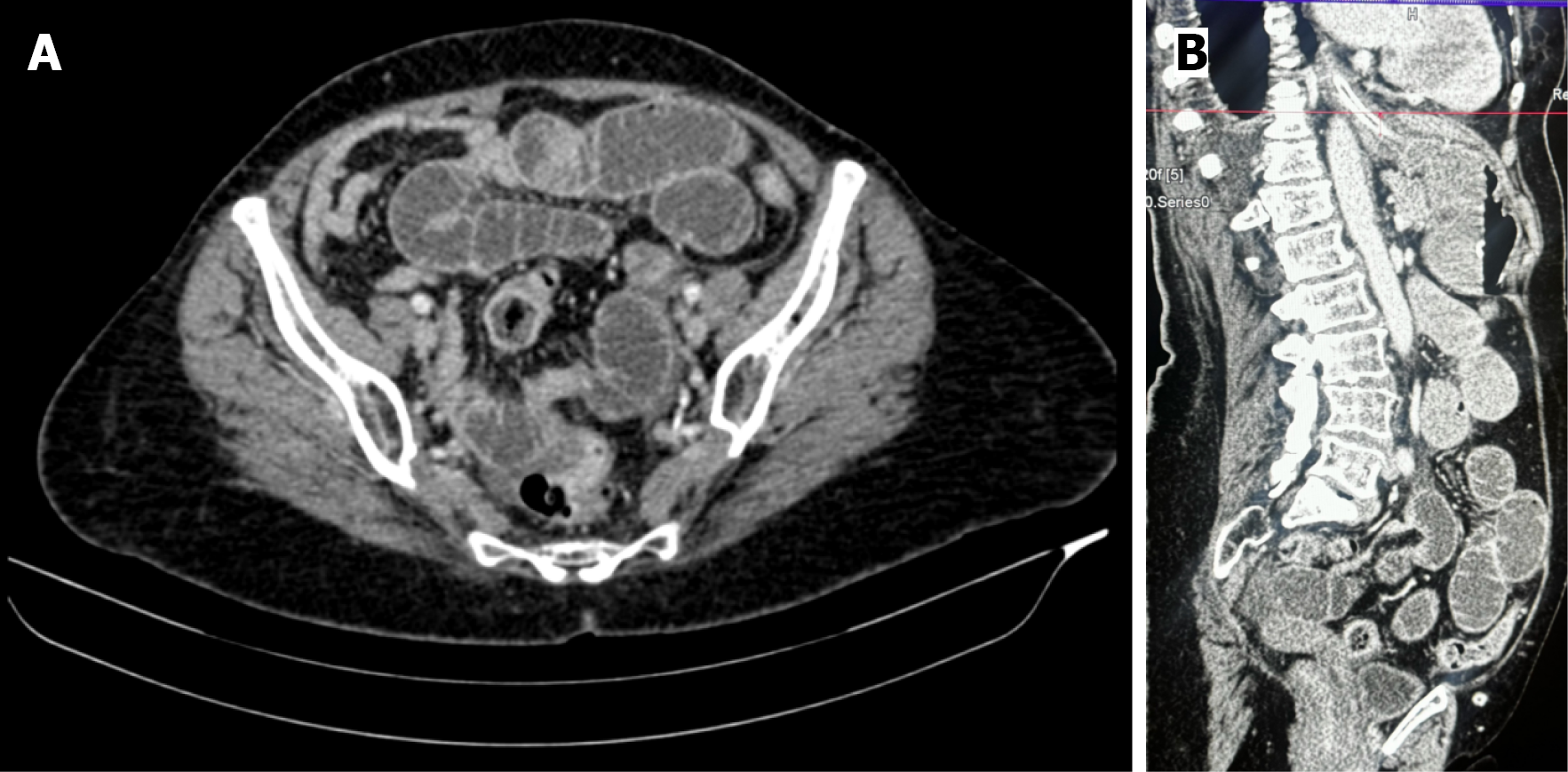
Figure 1 Contrast-enhanced computed tomography of the abdomen and pelvis.
A and B: Axial section shows multiple dilated, fluid-filled small bowel loops with preserved valvulae conniventes in the anterior and left mid-abdomen, consistent with proximal small bowel obstruction; an abrupt transition point is identified at a collapsed small bowel loop in the left paraumbilical region, beyond which the distal loops are decompressed.
- Citation: Joshi M, Jha S, Jha S, Jhawar N. Laparoscopic treatment of a transmesocolic internal hernia causing small bowel obstruction: A case report. World J Clin Cases 2026; 14(26): 126834
- URL: https://www.wjgnet.com/2307-8960/full/v14/i26/126834.htm
- DOI: https://dx.doi.org/10.12998/wjcc.126834