
















Copyright: ©Author(s) 2026.
World J Clin Cases. Sep 16, 2026; 14(26): 126833
Published online Sep 16, 2026. doi: 10.12998/wjcc.126833
Published online Sep 16, 2026. doi: 10.12998/wjcc.126833
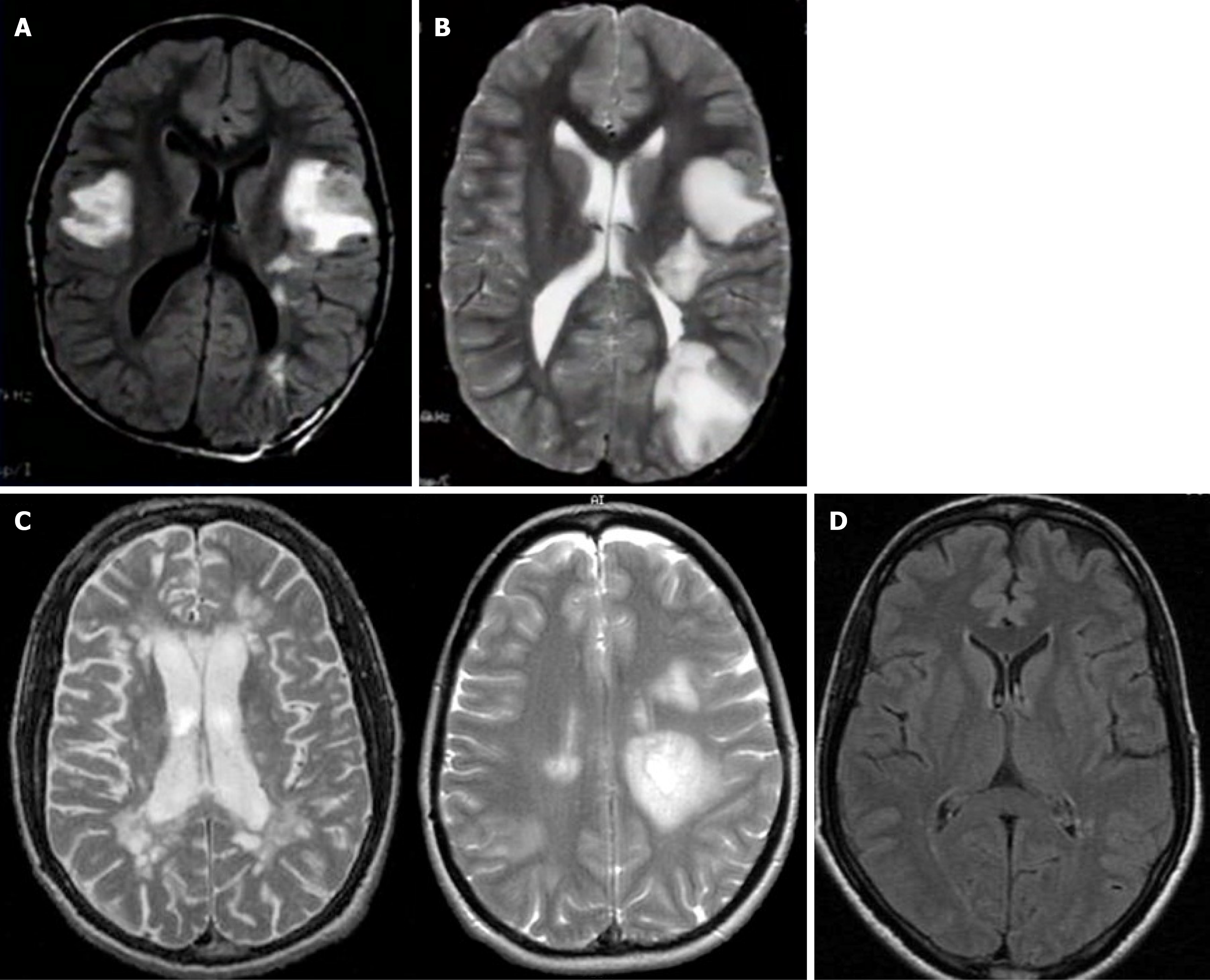
Figure 4 Case 2.
A and B: Brain magnetic resonance imaging (MRI) of case 2 at initial acute presentation (day 0). Axial T2-weighted fluid-attenuated inversion recovery (FLAIR) sequence (A) and axial T2-weighted fast spin-echo sequence (B), both demonstrating large, confluent, poorly marginated bilateral cerebral white-matter hyperintensities consistent with acute disseminated encephalomyelitis, with no restricted diffusion or pathological contrast enhancement; C: Pre-retreatment follow-up brain MRI of case 2 obtained at 3-month readmission (day +90, before corticosteroid retreatment). Showing persistent white-matter hyperintensities without new demyelinating lesions; D: Final follow-up axial T2-weighted FLAIR brain MRI of case 2 obtained at month 4 (day +120, exactly 4 weeks following the completion of repeat corticosteroid therapy). Demonstrating near-complete resolution of the prior cerebral white-matter lesions and absence of new demyelinating activity.
- Citation: Al-Beltagi M, Nazeer JA, Elbeltagi RM. Contrasting diagnostic challenges of headache before and after acute disseminated encephalomyelitis in children: Two case reports and review of literature. World J Clin Cases 2026; 14(26): 126833
- URL: https://www.wjgnet.com/2307-8960/full/v14/i26/126833.htm
- DOI: https://dx.doi.org/10.12998/wjcc.126833