
















Copyright: ©Author(s) 2026.
World J Clin Cases. Sep 16, 2026; 14(26): 126833
Published online Sep 16, 2026. doi: 10.12998/wjcc.126833
Published online Sep 16, 2026. doi: 10.12998/wjcc.126833
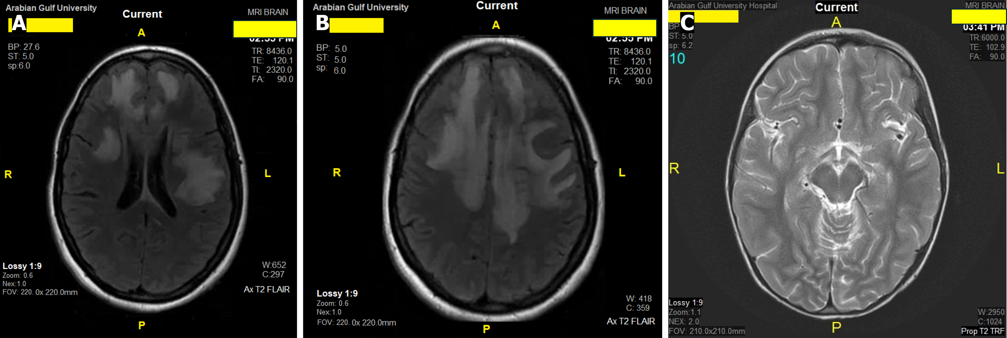
Figure 3 Case 1.
A and B: Axial T2-weighted fluid-attenuated inversion recovery brain magnetic resonance imaging (MRI) of case 1 obtained during initial acute presentation (day +3). Demonstrating multiple large, asymmetric white-matter lesions with poorly defined margins and relative periventricular sparing, accompanied by bilateral fairly symmetric hyperintensities involving the basal ganglia (more prominent on the left) with mild mass effect and no restricted diffusion. Institutional metadata has been de-identified in accordance with privacy guidelines; C: Axial T2-weighted brain MRI of case 1 obtained at 1-month follow-up (day +30). Demonstrating marked interval radiological improvement and near-complete resolution of the previously observed cerebral white-matter and basal ganglia abnormalities. Institutional metadata has been de-identified. FLAIR: Fluid-attenuated inversion recovery.
- Citation: Al-Beltagi M, Nazeer JA, Elbeltagi RM. Contrasting diagnostic challenges of headache before and after acute disseminated encephalomyelitis in children: Two case reports and review of literature. World J Clin Cases 2026; 14(26): 126833
- URL: https://www.wjgnet.com/2307-8960/full/v14/i26/126833.htm
- DOI: https://dx.doi.org/10.12998/wjcc.126833