
















Copyright: ©Author(s) 2026.
World J Clin Cases. Sep 16, 2026; 14(26): 126744
Published online Sep 16, 2026. doi: 10.12998/wjcc.126744
Published online Sep 16, 2026. doi: 10.12998/wjcc.126744
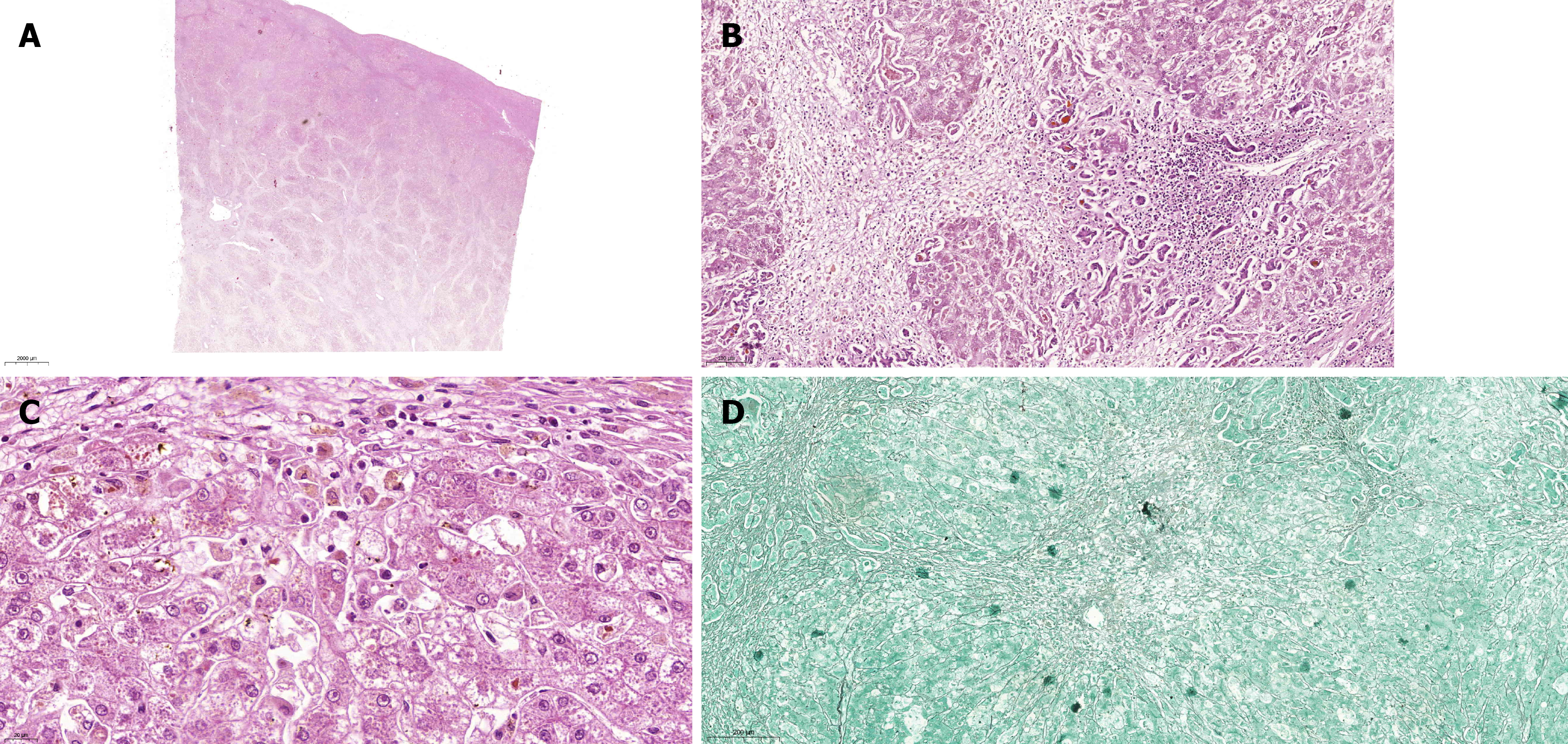
Figure 3 Histopathological examination of the explanted native liver.
A: Low-power panoramic view showing extensive confluent areas of hepatic parenchymal necrosis with marked architectural distortion, interspersed with relatively preserved/regenerative parenchyma [hematoxylin and eosin (H&E), digital magnification 0.6×; scale bar = 2000 μm]; B: Intermediate-power view showing extensive hepatocellular necrosis with marked architectural disruption, regenerative changes, focal inflammatory cell aggregates, and bile plugs consistent with cholestasis (H&E, digital magnification 10.9×; scale bar = 100 μm); C: High-power view demonstrating regenerative hepatocellular changes with enlarged hepatocytes and nuclear variability, associated with prominent canalicular and intracellular bile pigment consistent with marked cholestasis (H&E, digital magnification 44.4×; scale bar = 20 μm); D: Reticulin stain demonstrating marked disruption and collapse of the reticulin framework in areas of severe parenchymal injury (digital magnification 8.0×; scale bar = 200 μm). These histopathological findings were nonspecific and did not establish the etiology of the acute liver injury.
- Citation: Ponte RV, Faria AA, Guedes LR, Yamashiro IS, Macedo ML, Lima MC, Souza PF, Vidigal PV, Penna FG, Sancio JB. Acute liver failure temporally associated with intensive consumption of Plantago major tea: A case report. World J Clin Cases 2026; 14(26): 126744
- URL: https://www.wjgnet.com/2307-8960/full/v14/i26/126744.htm
- DOI: https://dx.doi.org/10.12998/wjcc.126744