
















Copyright: ©Author(s) 2026.
World J Clin Cases. Sep 16, 2026; 14(26): 126222
Published online Sep 16, 2026. doi: 10.12998/wjcc.126222
Published online Sep 16, 2026. doi: 10.12998/wjcc.126222
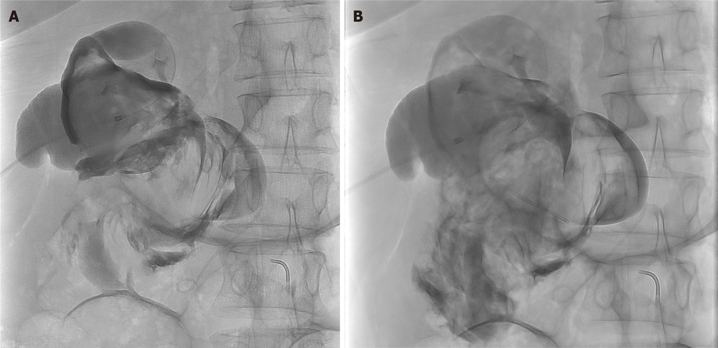
Figure 4 Cholecystostomy cholangiogram showing common bile duct extravasation.
Fluoroscopic images obtained via injection of contrast through percutaneously placed cholecystostomy tube. A: Early-phase image showing opacification of the gallbladder and proximal biliary tree, confirming the appropriate position of the tube; B: Progressive-phase image obtained with continued contrast injection, demonstrating extravasation of the contrast into an irregular, poorly marginated collection consistent with perforation of the bile duct and active bile leak.
- Citation: Bautista LYA, Lee M, Devgan K, Longo J, Singh S, Bakdash K, Ahmed M, Agrawal R. Preoperative cholangioscopic identification of traumatic type IVa choledochal cyst perforation after motor vehicle collision: A case report. World J Clin Cases 2026; 14(26): 126222
- URL: https://www.wjgnet.com/2307-8960/full/v14/i26/126222.htm
- DOI: https://dx.doi.org/10.12998/wjcc.126222