
















Copyright: ©Author(s) 2026.
World J Clin Cases. Sep 16, 2026; 14(26): 125200
Published online Sep 16, 2026. doi: 10.12998/wjcc.125200
Published online Sep 16, 2026. doi: 10.12998/wjcc.125200
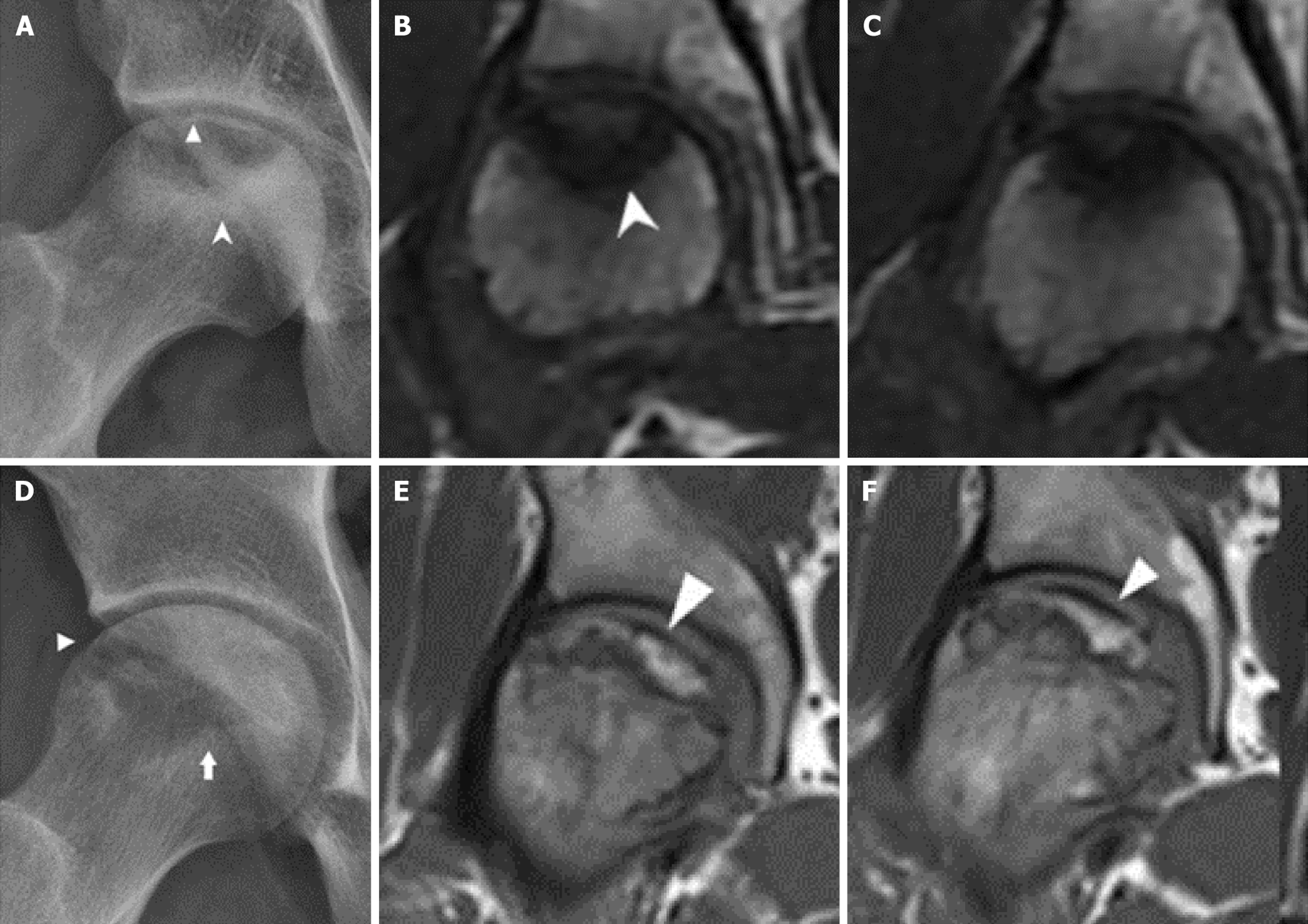
Figure 2 Representative radiographs and T1-weighted magnetic resonance images of subchondral stress fracture of the femoral head and osteonecrosis of the femoral head.
A-F: Imaging findings in a 20-year-old male recruit with subchondral stress fracture of the femoral head (SSFFH) (A-C) and a 45-year-old military officer with osteonecrosis of the femoral head (ONFH) (D-F). A and D: Radiographs show similar articular-surface collapse in both SSFFH and ONFH; the white triangles indicate subchondral fracture; B and C: Magnetic resonance imaging (MRI) of SSFFH reveals an irregular subchondral low-signal band (white arrow) with surrounding bone marrow edema and an irregular, thickened sclerotic margin; E and F: MRI of ONFH demonstrates a smoother, serpiginous low-signal band demarcating the necrotic segment, with a subchondral fracture indicated by the white triangle. These findings illustrate the potential overlap on radiographs and the added value of MRI for differential classification.
- Citation: Jiang YF, Wang JB, Zhao B. Subchondral stress fractures of the femoral head associated with military training: Clinical features and analysis. World J Clin Cases 2026; 14(26): 125200
- URL: https://www.wjgnet.com/2307-8960/full/v14/i26/125200.htm
- DOI: https://dx.doi.org/10.12998/wjcc.125200