















Copyright: ©Author(s) 2026.
World J Clin Cases. Sep 6, 2026; 14(25): 124535
Published online Sep 6, 2026. doi: 10.12998/wjcc.124535
Published online Sep 6, 2026. doi: 10.12998/wjcc.124535
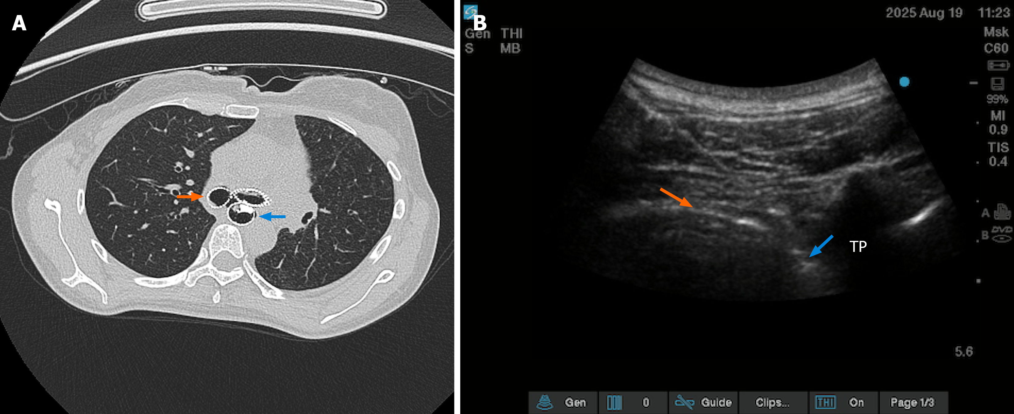
Figure 1 Computed tomography and ultrasound images.
A: Showed dual stents and the esophageal cancer lesion orange arrow shows the tracheal stent; blue arrow shows the esophageal stent; B: Downward of the pleura after thoracic paravertebral block orange arrow shows the pleura; blue arrow shows the paravertebral space. TP: Transverse process.
- Citation: Tan RX, Liu HB, Zuo YD, Qiu YH. Monitored anesthesia care combined with thoracic paravertebral block for percutaneous endoscopic gastrostomy: A case report. World J Clin Cases 2026; 14(25): 124535
- URL: https://www.wjgnet.com/2307-8960/full/v14/i25/124535.htm
- DOI: https://dx.doi.org/10.12998/wjcc.124535