
















Copyright: ©Author(s) 2026.
World J Clin Cases. Aug 26, 2026; 14(24): 123416
Published online Aug 26, 2026. doi: 10.12998/wjcc.123416
Published online Aug 26, 2026. doi: 10.12998/wjcc.123416
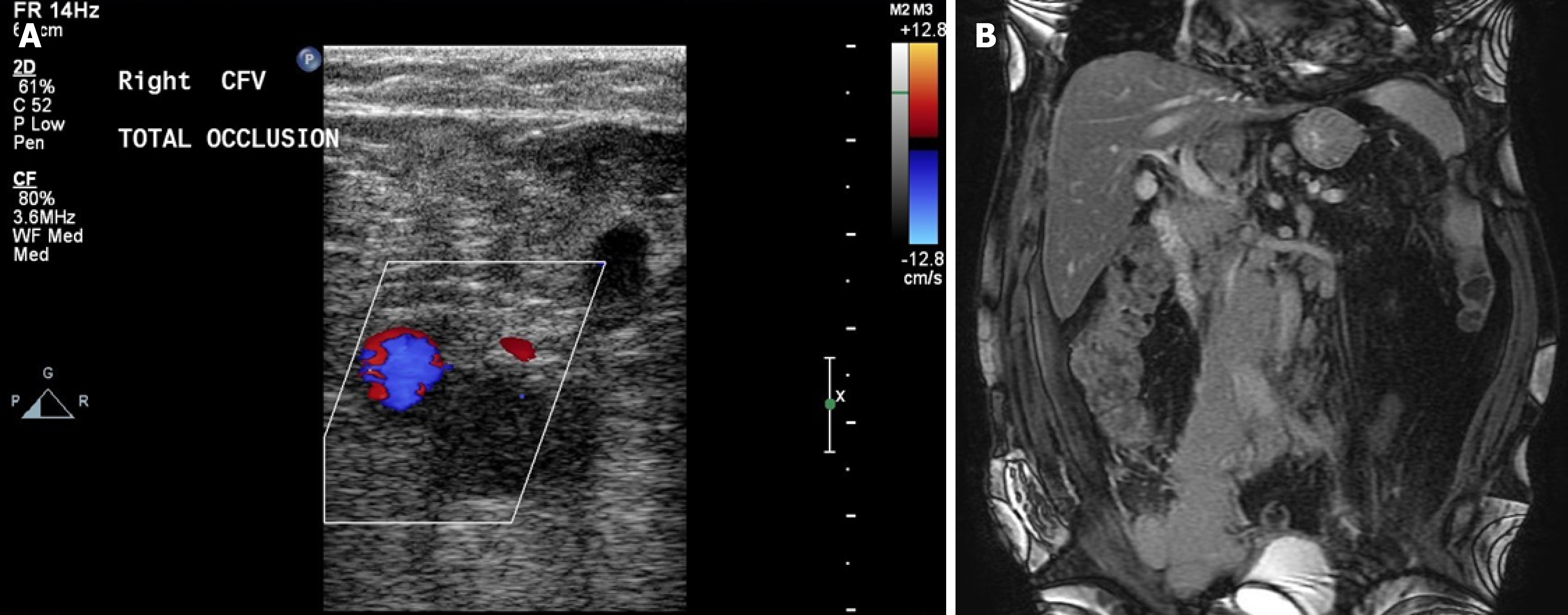
Figure 1 Imaging examinations.
A: Doppler showed total occlusion of the right common femoral vein; B: Magnetic resonance imaging showed a huge mass over the paraaortic, paraspinal and right pelvic region encasing abdominal aorta, inferior vena cava, right iliac artery and vein.
- Citation: Chen TC, Hsu JF. Lymphoma-associated deep vein thrombosis mimicking cellulitis in an elderly patient with diabetes: A case report. World J Clin Cases 2026; 14(24): 123416
- URL: https://www.wjgnet.com/2307-8960/full/v14/i24/123416.htm
- DOI: https://dx.doi.org/10.12998/wjcc.123416