
















Copyright: ©Author(s) 2026.
World J Clin Cases. Aug 26, 2026; 14(24): 123303
Published online Aug 26, 2026. doi: 10.12998/wjcc.123303
Published online Aug 26, 2026. doi: 10.12998/wjcc.123303
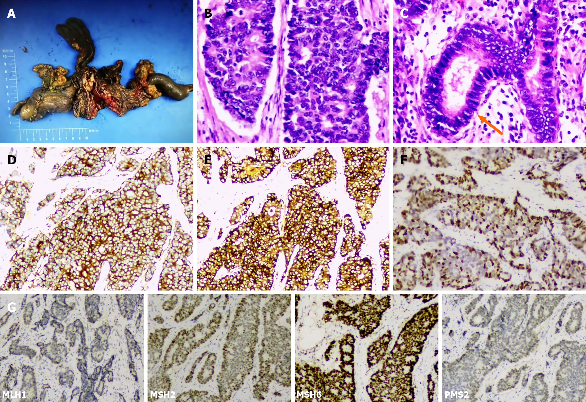
Figure 2 Histopathological examination of the resected specimen.
A: Gross appearance. The resected specimen revealed a nodular, protuberant mass in the distal common bile duct measuring 2 cm × 1.5 cm × 1 cm, located 0.6 cm from the cystic duct confluence and 5.5 cm from the duodenal papilla. The cut surface was grayish-white and firm in consistency; B: Microscopic findings (hematoxylin and eosin staining, × 200) – the tumor was composed of large cells arranged in nests and sheets. The tumor cells exhibited abundant cytoplasm, coarse chromatin, prominent nucleoli, and brisk mitotic activity (> 10/high-power field); C: Microscopic findings (hematoxylin and eosin staining, × 200) - a focal moderately differentiated adenocarcinoma component is identified, accounting for approximately 10% of the tumor volume; D: Immunohistochemistry (× 200) showing diffuse cytoplasmic positivity for synaptophysin; E: Immunohistochemistry (× 200) showing focal cytoplasmic positivity for chromogranin A (CgA); F: Ki-67 immunohistochemistry (× 200) showing a proliferation index of approximately 60%; G: Mismatch repair (MMR) protein immunohistochemistry (× 200) showing retained nuclear expression of MLH1, MSH2, MSH6, and PMS2, consistent with a proficient MMR phenotype.
- Citation: Zheng BQ, Wang ZP, Feng RZ, Zheng ZC, Wang HJ, Wang H, Tuo HF. Large cell neuroendocrine carcinoma of the distal common bile duct: A case report and literature review. World J Clin Cases 2026; 14(24): 123303
- URL: https://www.wjgnet.com/2307-8960/full/v14/i24/123303.htm
- DOI: https://dx.doi.org/10.12998/wjcc.123303