
















Copyright: ©Author(s) 2026.
World J Clin Cases. Aug 26, 2026; 14(24): 123303
Published online Aug 26, 2026. doi: 10.12998/wjcc.123303
Published online Aug 26, 2026. doi: 10.12998/wjcc.123303
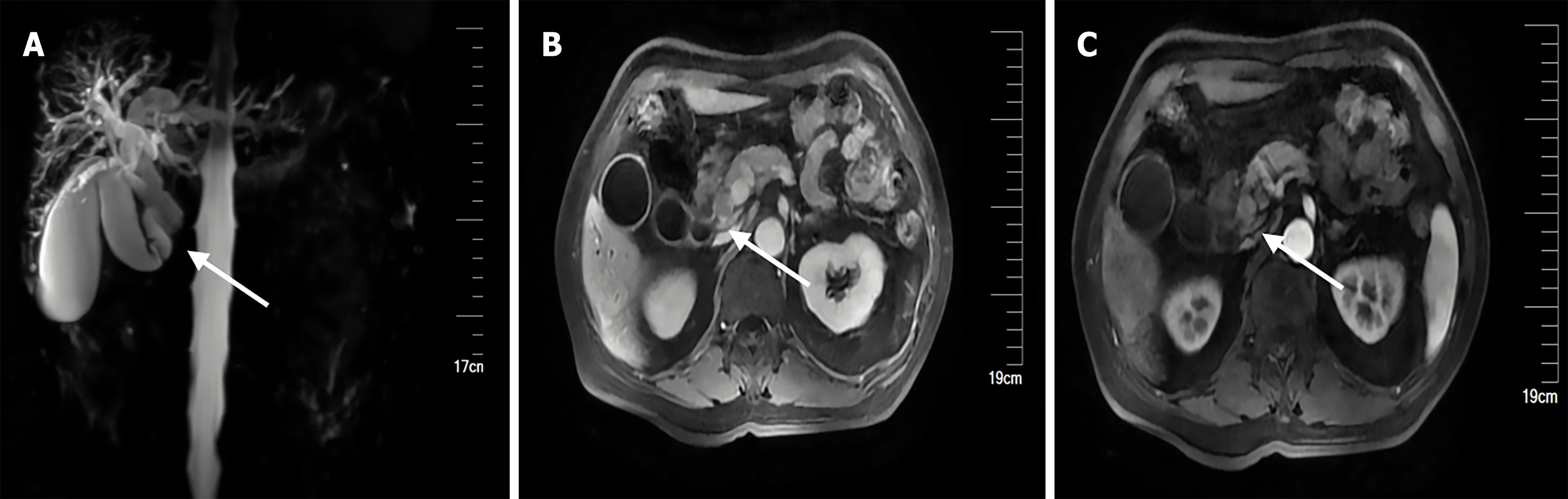
Figure 1 Magnetic resonance image.
A: Magnetic resonance cholangiopancreatography showed segmental interruption of the middle and distal common bile duct (arrow) with marked dilatation of the upstream intrahepatic and extrahepatic bile ducts; B: Contrast-enhanced magnetic resonance imaging in the arterial phase reveals a heterogeneous enhancing nodular mass measuring approximately 16 mm × 14 mm in the distal common bile duct (arrow); C: Portal venous/delayed phase demonstrates rapid washout of the mass.
- Citation: Zheng BQ, Wang ZP, Feng RZ, Zheng ZC, Wang HJ, Wang H, Tuo HF. Large cell neuroendocrine carcinoma of the distal common bile duct: A case report and literature review. World J Clin Cases 2026; 14(24): 123303
- URL: https://www.wjgnet.com/2307-8960/full/v14/i24/123303.htm
- DOI: https://dx.doi.org/10.12998/wjcc.123303