
















Copyright: ©Author(s) 2026.
World J Clin Cases. Aug 16, 2026; 14(23): 121367
Published online Aug 16, 2026. doi: 10.12998/wjcc.121367
Published online Aug 16, 2026. doi: 10.12998/wjcc.121367
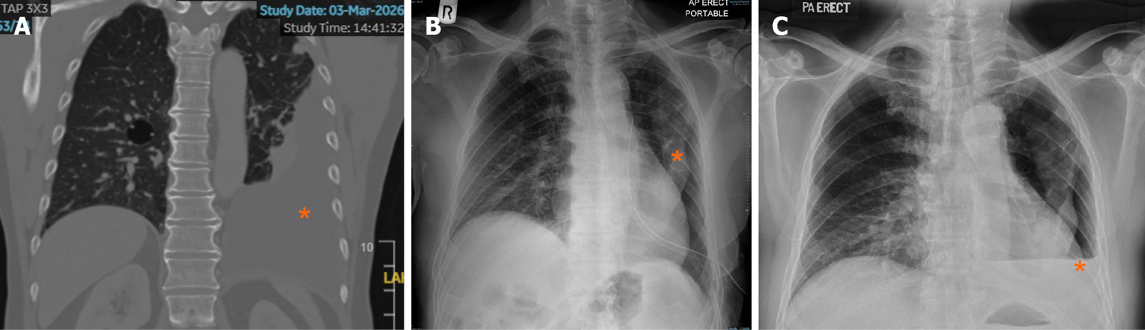
Figure 1 Radiology images.
A: Pre-procedural computed tomography scan image on 3 March 2026, showing a moderately large left-sided pleural effusion (asterisk); B: Post-procedure chest X-ray image on March 5, 2026, 2 hours after thoracoscopy, showing complete pleural fluid drainage, a left-sided chest tube in situ, and an unexpanded left lung (asterisk); C: Post-procedure chest X-ray image on March 20, 2026, 2 weeks after thoracoscopy, showing partial accumulation of pleural fluid, an air-fluid level (asterisk), and an unexpandable left lung.
- Citation: See KC. Performing medical thoracoscopy safely in the outpatient setting: A case report. World J Clin Cases 2026; 14(23): 121367
- URL: https://www.wjgnet.com/2307-8960/full/v14/i23/121367.htm
- DOI: https://dx.doi.org/10.12998/wjcc.121367