
















Copyright: ©Author(s) 2026.
World J Clin Cases. Aug 6, 2026; 14(22): 122012
Published online Aug 6, 2026. doi: 10.12998/wjcc.122012
Published online Aug 6, 2026. doi: 10.12998/wjcc.122012
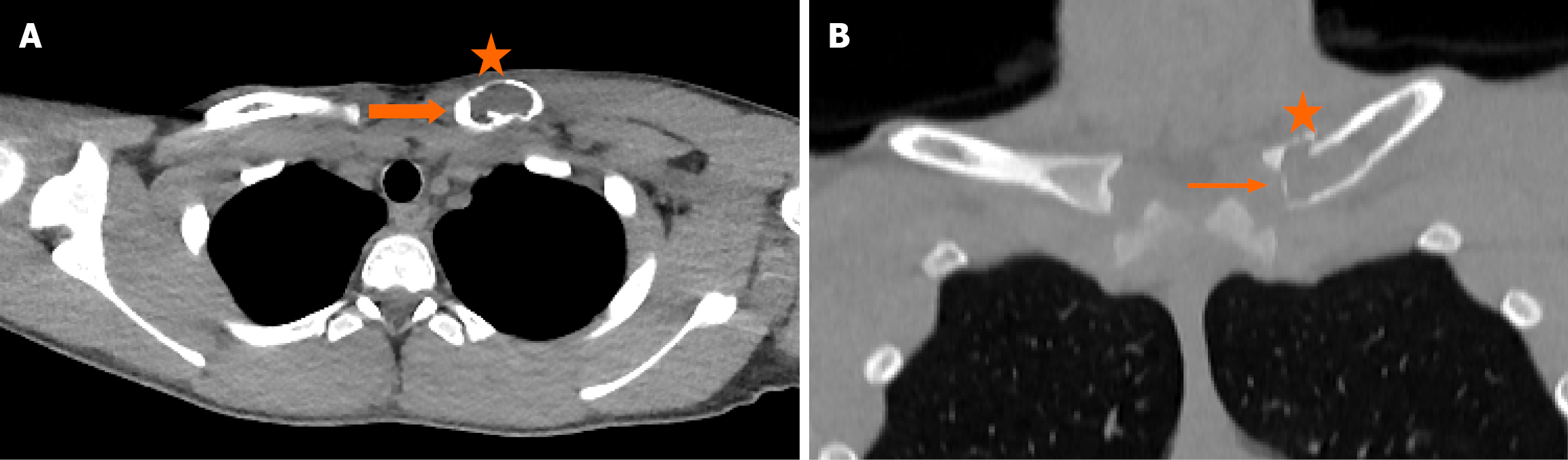
Figure 1 Axial computed tomography and coronal computed tomography chest.
A: Axial computed tomography (CT) chest shows a lytic lesion (asterisk, 4 cm × 2 cm) that affects the medial aspect of the left clavicle with cortical diaphyseal expansion (thick arrow) reaching the inner surface of the sternoclavicular joint; B: Coronal CT chest shows perilesional cortical thinning (asterisk) with focal cortical disruption (slim arrow) and mild periosteal reaction.
- Citation: Gkougkoulias AN, Vlychou M, Dailiana ZH. Monostotic chronic nonbacterial osteomyelitis/chronic recurrent multifocal osteomyelitis of clavicle mimicking malignant bone tumor: A case report and review of literature. World J Clin Cases 2026; 14(22): 122012
- URL: https://www.wjgnet.com/2307-8960/full/v14/i22/122012.htm
- DOI: https://dx.doi.org/10.12998/wjcc.122012