
















Copyright: ©Author(s) 2026.
World J Clin Cases. Aug 6, 2026; 14(22): 121636
Published online Aug 6, 2026. doi: 10.12998/wjcc.121636
Published online Aug 6, 2026. doi: 10.12998/wjcc.121636
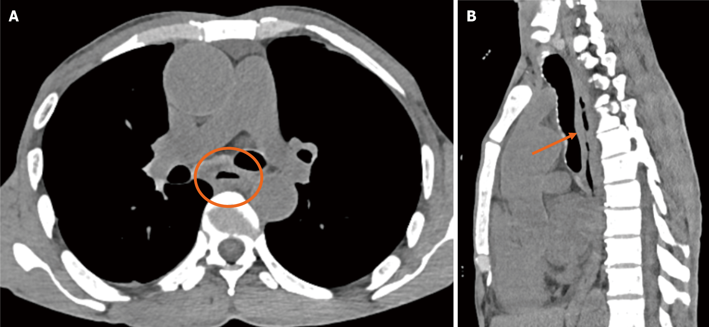
Figure 1 Contrast-enhanced computed tomography chest showing diffuse oesophageal thickening at mid esophageal level (orange circle in A and orange arrow in B).
No definite foreign or pneumomediastinum was identified. A: Cross-sectional image; B: Axial image.
- Citation: Reddy RT, Vaithiyam V, Hussain GZ, Arul Y, Sonika U, Dalal A, Srivastava S, Sachdeva S. Successful endoscopic fragmentation and removal of an impacted esophageal denture: A case report. World J Clin Cases 2026; 14(22): 121636
- URL: https://www.wjgnet.com/2307-8960/full/v14/i22/121636.htm
- DOI: https://dx.doi.org/10.12998/wjcc.121636