
















Copyright: ©Author(s) 2026.
World J Clin Cases. Jul 16, 2026; 14(20): 121466
Published online Jul 16, 2026. doi: 10.12998/wjcc.121466
Published online Jul 16, 2026. doi: 10.12998/wjcc.121466
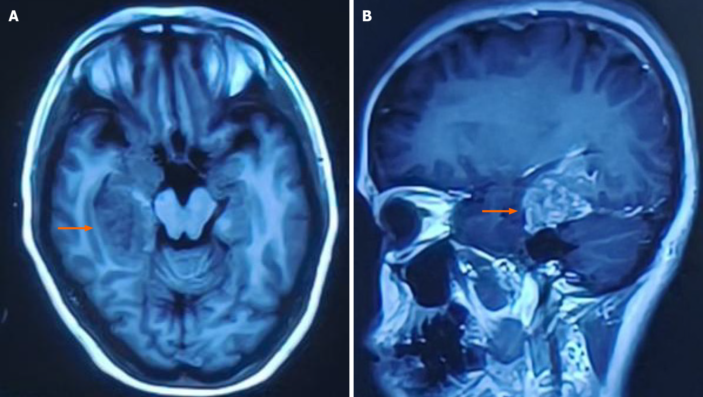
Figure 4 Cranial magnetic resonance imaging showing a T1 hypointense area in the right thalamus (orange arrow).
A: Axial section; B: Sagittal section [provided by Huashan Hospital (West Campus), affiliated to Fudan University, Shanghai, China].
- Citation: Xu KC, Kong XF, Qian W, Liang B, Li DG, Korpan NN. Long-term survival in patients with primary malignant brain tumors after postoperative home hydrogen inhalation: Three case reports. World J Clin Cases 2026; 14(20): 121466
- URL: https://www.wjgnet.com/2307-8960/full/v14/i20/121466.htm
- DOI: https://dx.doi.org/10.12998/wjcc.121466