
















©The Author(s) 2026.
World J Clin Cases. Jan 16, 2026; 14(2): 116519
Published online Jan 16, 2026. doi: 10.12998/wjcc.v14.i2.116519
Published online Jan 16, 2026. doi: 10.12998/wjcc.v14.i2.116519
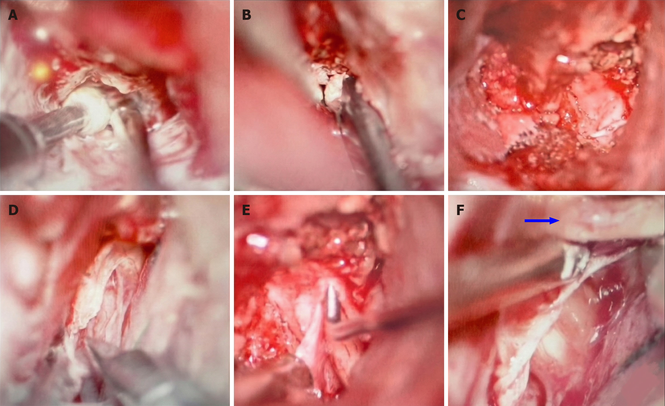
Figure 3 Key surgical steps in extradural anterior clinoidectomy and optic nerve decompression.
A: Extradural drilling of the anterior clinoid process using a diamond high-speed drill; B: Opening the optic canal with 1-mm Kerrison punch; C: Demonstration of extradural clinoidectomy and decompression of the optic canal; D: Opening the optic nerve dural sleeve; E: Use of a mini hook to verify complete optic nerve decompression intradural; F: Completely decompressed and free optic nerve, with adipose tissues (indicated by blue arrow) cleared away.
- Citation: Tlaiss Y, Abou Zeki L, Farhat H, Warrak J, Yazbeck M, Al-Awar O. Intraosseous lipoma of the sphenoid bone causing optic nerve compression: A case report. World J Clin Cases 2026; 14(2): 116519
- URL: https://www.wjgnet.com/2307-8960/full/v14/i2/116519.htm
- DOI: https://dx.doi.org/10.12998/wjcc.v14.i2.116519