
















©The Author(s) 2026.
World J Clin Cases. Jan 16, 2026; 14(2): 116519
Published online Jan 16, 2026. doi: 10.12998/wjcc.v14.i2.116519
Published online Jan 16, 2026. doi: 10.12998/wjcc.v14.i2.116519
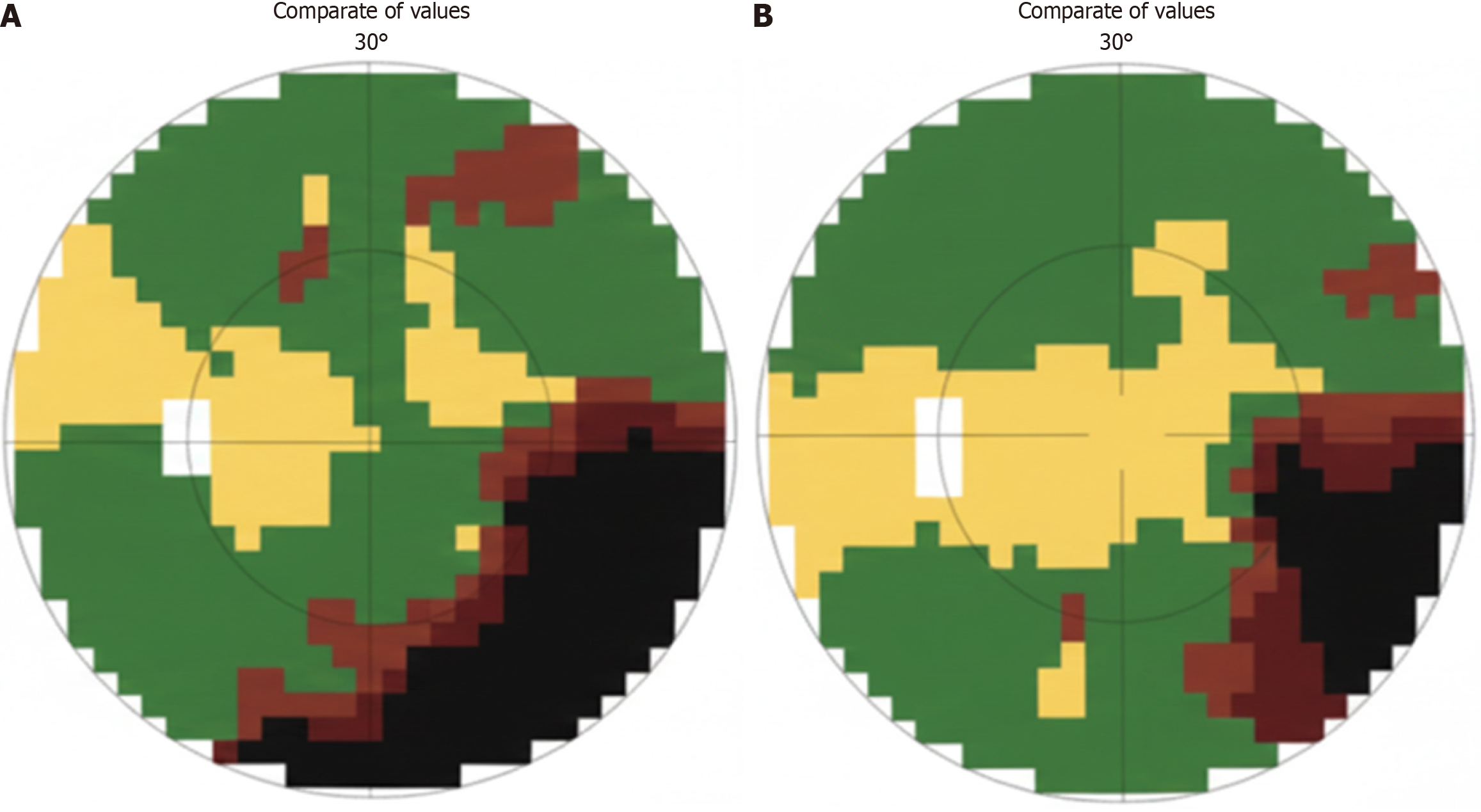
Figure 1 Visual field perimetry (octopus 101) demonstrating the preoperative and postoperative status.
A: Left eye (Oculus Sinister) preoperatively, exhibiting significant visual field loss, particularly affecting the inferotemporal region; B: Left eye (Oculus Sinister) postoperatively, revealing marked improvement and substantial recovery of visual fields after surgical decompression.
- Citation: Tlaiss Y, Abou Zeki L, Farhat H, Warrak J, Yazbeck M, Al-Awar O. Intraosseous lipoma of the sphenoid bone causing optic nerve compression: A case report. World J Clin Cases 2026; 14(2): 116519
- URL: https://www.wjgnet.com/2307-8960/full/v14/i2/116519.htm
- DOI: https://dx.doi.org/10.12998/wjcc.v14.i2.116519