
















©The Author(s) 2026.
World J Clin Cases. Jan 16, 2026; 14(2): 114671
Published online Jan 16, 2026. doi: 10.12998/wjcc.v14.i2.114671
Published online Jan 16, 2026. doi: 10.12998/wjcc.v14.i2.114671
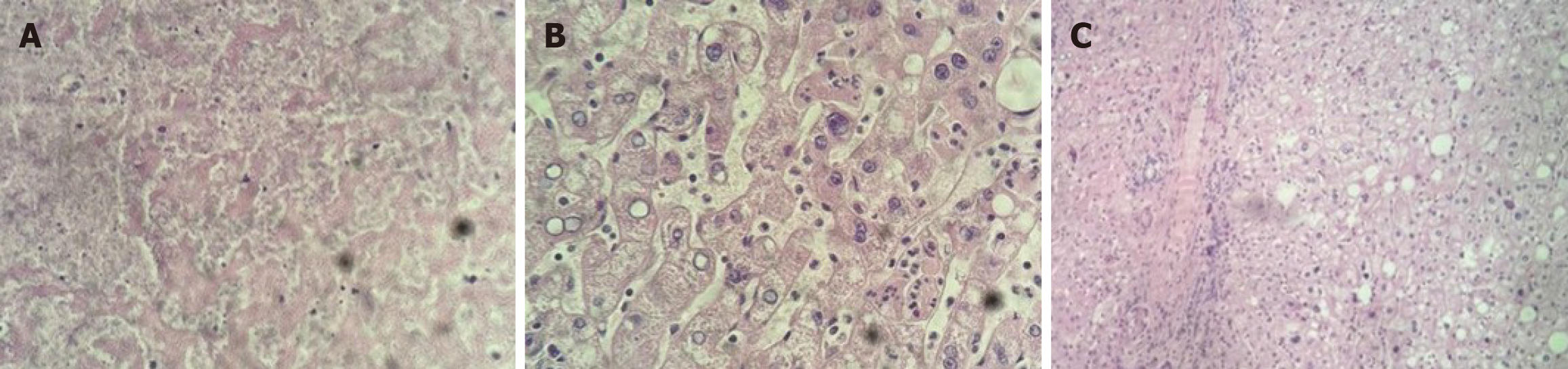
Figure 2 Histopathological and immunohistochemical examination of the resected specimen.
A: Area of coagulative necrosis within the adenoma [hematoxylin and eosin (H&E), 100 ×]; B: Higher magnification showing benign hepatocytes arranged in plates without portal tracts (H&E, 400 ×); C: Lower magnification showing preserved hepatic parenchyma adjacent to the lesion (H&E, 100 ×).
- Citation: Pagani Junior MA, Siman Lopes L, Sergi Filho FA, Goes Scorsioni FA, Tavares Filho MB, Maluf Amaral RP, Medeiros LE, Guillen Carneiro LI, Belluzzo HG, Vinholo TR, Buchaim DV, Buchaim RL. Partial left hepatectomy in a young patient with ruptured hepatic adenoma: A case report. World J Clin Cases 2026; 14(2): 114671
- URL: https://www.wjgnet.com/2307-8960/full/v14/i2/114671.htm
- DOI: https://dx.doi.org/10.12998/wjcc.v14.i2.114671