
















©The Author(s) 2026.
World J Clin Cases. Jan 16, 2026; 14(2): 114671
Published online Jan 16, 2026. doi: 10.12998/wjcc.v14.i2.114671
Published online Jan 16, 2026. doi: 10.12998/wjcc.v14.i2.114671
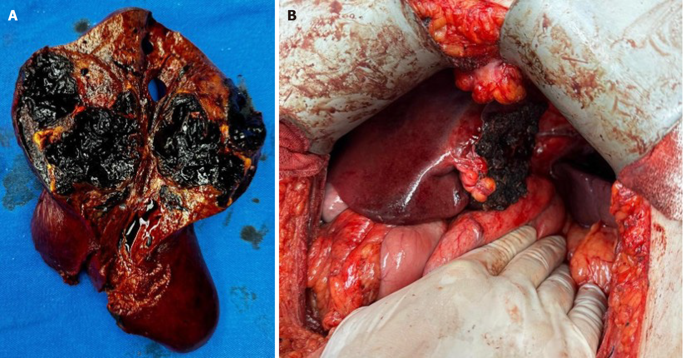
Figure 1 Imaging and macroscopic examination of the hepatic adenoma.
A: Surgical specimen showing a well-defined and partially encapsulated subcapsular lesion in the left hepatic lobe; B: Intraoperative view after resection of the hepatic lesion, showing exposure of the remaining liver parenchyma.
- Citation: Pagani Junior MA, Siman Lopes L, Sergi Filho FA, Goes Scorsioni FA, Tavares Filho MB, Maluf Amaral RP, Medeiros LE, Guillen Carneiro LI, Belluzzo HG, Vinholo TR, Buchaim DV, Buchaim RL. Partial left hepatectomy in a young patient with ruptured hepatic adenoma: A case report. World J Clin Cases 2026; 14(2): 114671
- URL: https://www.wjgnet.com/2307-8960/full/v14/i2/114671.htm
- DOI: https://dx.doi.org/10.12998/wjcc.v14.i2.114671