
















Copyright: ©Author(s) 2026.
World J Clin Cases. Jun 26, 2026; 14(18): 121564
Published online Jun 26, 2026. doi: 10.12998/wjcc.121564
Published online Jun 26, 2026. doi: 10.12998/wjcc.121564
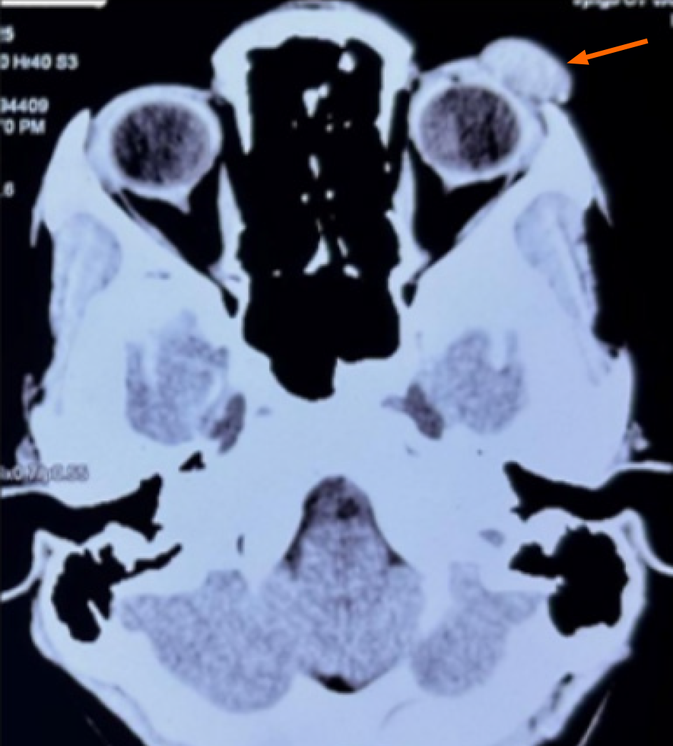
Figure 3 Computed tomography of the orbits showing left upper eyelid lesion.
Axial non-contrast computed tomography image demonstrating a well-defined soft tissue lesion in the left periorbital region (orange arrow) involving the upper eyelid and supraorbital area without evidence of underlying bony erosion or intraorbital extension. The imaging features were suggestive of a benign cystic lesion, such as an epidermal inclusion cyst.
- Citation: Panda BB, Gokhale S, Mishra P, Sethy M. Primary arteriovenous malformation of the eyelid mimicking a dermoid cyst: A case report. World J Clin Cases 2026; 14(18): 121564
- URL: https://www.wjgnet.com/2307-8960/full/v14/i18/121564.htm
- DOI: https://dx.doi.org/10.12998/wjcc.121564