
















Copyright: ©Author(s) 2026.
World J Clin Cases. Jun 26, 2026; 14(18): 121564
Published online Jun 26, 2026. doi: 10.12998/wjcc.121564
Published online Jun 26, 2026. doi: 10.12998/wjcc.121564
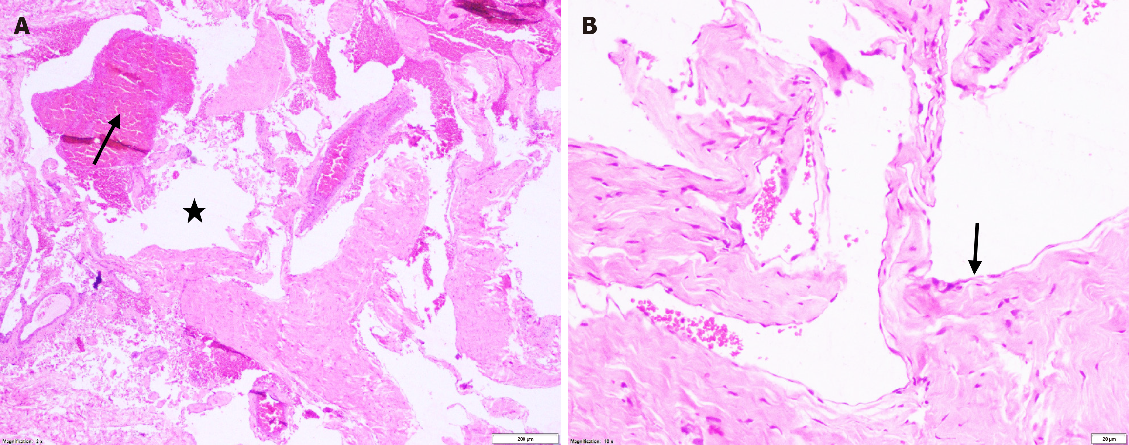
Figure 2 Histopathological features of the lesion (hematoxylin and eosin staining).
A: Low-power view showing interconnected congested veins (black arrow) and empty spaces i.e., lymphatics (shown by star) [hematoxylin and eosin (H&E), × 2] suggestive of a combined veno-lymphatic malformation; B: Higher magnification demonstrating irregular, tortuous vessels lined by a single layer of endothelial lining (black arrow) (H&E, × 10).
- Citation: Panda BB, Gokhale S, Mishra P, Sethy M. Primary arteriovenous malformation of the eyelid mimicking a dermoid cyst: A case report. World J Clin Cases 2026; 14(18): 121564
- URL: https://www.wjgnet.com/2307-8960/full/v14/i18/121564.htm
- DOI: https://dx.doi.org/10.12998/wjcc.121564