
















Copyright: ©Author(s) 2026.
World J Clin Cases. Jun 26, 2026; 14(18): 121564
Published online Jun 26, 2026. doi: 10.12998/wjcc.121564
Published online Jun 26, 2026. doi: 10.12998/wjcc.121564
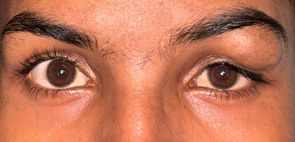
Figure 1 Preoperative clinical photograph showing left upper eyelid swelling.
Frontal view of the patient demonstrating a well-defined swelling in the temporal aspect of the left upper eyelid, with normal overlying skin and no visible vascular changes such as discoloration or dilated vessels. The contralateral eye appears normal.
- Citation: Panda BB, Gokhale S, Mishra P, Sethy M. Primary arteriovenous malformation of the eyelid mimicking a dermoid cyst: A case report. World J Clin Cases 2026; 14(18): 121564
- URL: https://www.wjgnet.com/2307-8960/full/v14/i18/121564.htm
- DOI: https://dx.doi.org/10.12998/wjcc.121564