
















Copyright: ©Author(s) 2026.
World J Clin Cases. Jun 26, 2026; 14(18): 121058
Published online Jun 26, 2026. doi: 10.12998/wjcc.121058
Published online Jun 26, 2026. doi: 10.12998/wjcc.121058
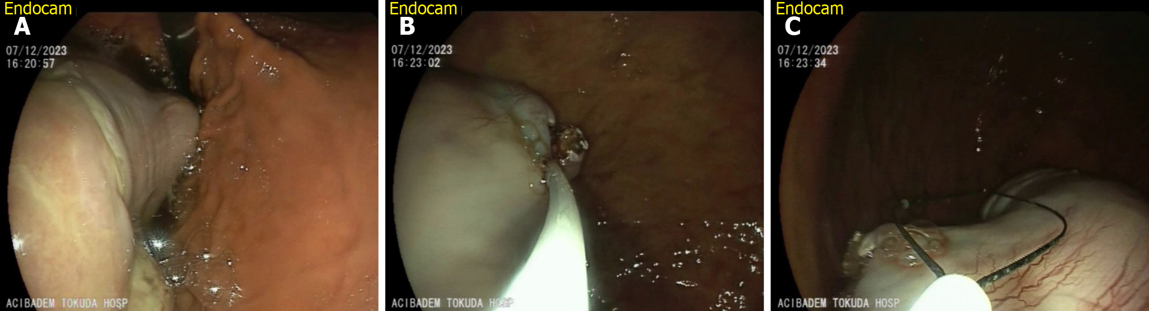
Figure 5 Endoscopic images of a giant fibrovascular polyp from esophagus into the gastric lumen.
A. Endoscopic view showing the giant fibrovascular polyp extending from the esophagus toward the gastric lumen; B: Close-up view of the polyp’s base and stalk entering the gastric lumen; C: Oblique view of the polyp showing the stalk’s attachment to the esophageal mucosa and spatial orientation within the lumen.
- Citation: Karagyozov P, Zhecheva I, Ivanov K, Galev I. Challenges in the diagnosis and treatment of giant fibrovascular polyps of the esophagus: Two case reports. World J Clin Cases 2026; 14(18): 121058
- URL: https://www.wjgnet.com/2307-8960/full/v14/i18/121058.htm
- DOI: https://dx.doi.org/10.12998/wjcc.121058