
















Copyright: ©Author(s) 2026.
World J Clin Cases. Jun 26, 2026; 14(18): 120279
Published online Jun 26, 2026. doi: 10.12998/wjcc.120279
Published online Jun 26, 2026. doi: 10.12998/wjcc.120279
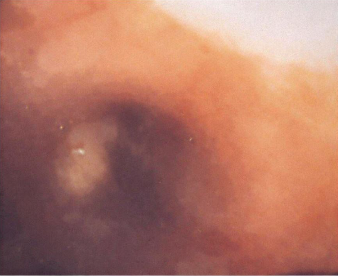
Figure 2 Bronchoscopic appearance of endobronchial chondroma.
Flexible bronchoscopy demonstrating a polypoid, smooth, vascularized endobronchial lesion partially obstructing the lumen of the left upper lobar bronchus. The mass appears broad-based, with intact overlying mucosa and no visible necrosis or ulceration, findings consistent with a benign endobronchial tumor subsequently confirmed as chondroma on histopathological examination.
- Citation: Dodos K, Kalamara TV, Georgakopoulou VE. Endobronchial chondroma presenting as recurrent pneumonia - diagnostic role of bronchoscopy and surgical management: A case report. World J Clin Cases 2026; 14(18): 120279
- URL: https://www.wjgnet.com/2307-8960/full/v14/i18/120279.htm
- DOI: https://dx.doi.org/10.12998/wjcc.120279